Rationale
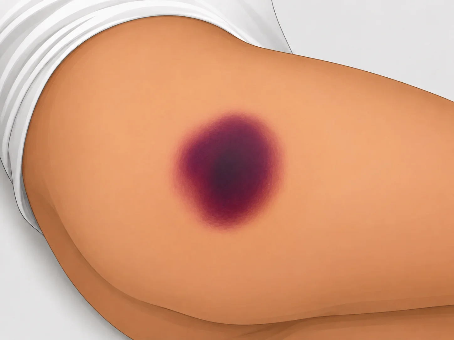
Putting the photograph and the description together, the skin surface is intact but discolored purple or maroon, and the area feels cooler and firmer than the surrounding tissue. This is the classic presentation of a
suspected deep tissue injury (sDTI), indicating that severe ischemic damage has already occurred in the soft tissue beneath the skin surface — that is, below the dermis. The correct answer is therefore
option 4.
Incorrect Options and Underlying Pathophysiology
Linking each option to its pathophysiologic mechanism clarifies why deep tissue injury, rather than another stage, must be suspected.
-
Option 1 (superficial stage 1 pressure injury): The hallmark of a stage 1 pressure injury is localized non-blanchable erythema with intact skin. Pressure damages dermal capillaries and produces reactive hyperemia, so the area typically feels warmer than surrounding tissue and may be edematous. In contrast, the lesion described is purple, cool, and firm, which points to deeper tissue ischemia and necrosis and distinguishes it from stage 1.
-
Options 2 and 5 (stage 3 and stage 4 pressure injuries): Stage 3 involves full-thickness skin loss with visible subcutaneous fat, and stage 4 involves exposure of fascia, muscle, or bone. Because the question states that "the skin is intact," neither stage 3 nor stage 4 — both of which require full-thickness skin loss — applies.
-
Option 3 (unstageable pressure injury): An unstageable injury is one in which eschar or yellow slough obscures the wound bed so that the depth of tissue damage cannot be visually determined. In this lesion the skin remains closed and only discoloration is present; no slough or eschar covers a wound bed, so it is not unstageable.
-
Option 4 (suspected deep tissue injury): This correct answer results when intense, sustained pressure or shear force directly damages muscle and soft tissue overlying a bony prominence. Muscle tissue is far more vulnerable to ischemia than skin, so extensive ischemic necrosis can already be under way in the muscle layer while the skin surface still looks intact. As vessels are destroyed, blood components leak into the tissue and produce purple discoloration at the surface; interrupted blood flow makes the tissue feel cool, and edema and induration make it feel firm. These findings reflect serious injury to the soft tissue below the dermis rather than superficial skin damage.
Clinical Application and Assessment
Early detection of this kind of deep tissue damage is critical, because visual inspection alone identifies only damage that has already progressed. The supporting evidence
[1] shows that measuring changes in subepidermal moisture (SEM) can predict early tissue damage before the skin becomes erythematous or progresses to a stage 1 pressure injury. In that study, rising SEM values reflected localized inflammation and edema appearing roughly one week before skin damage became visible. In practice, therefore, when signs of sDTI such as purple discoloration are observed, they should be treated not as simple skin discoloration but as evidence of established deep tissue damage, prompting immediate offloading interventions and close monitoring for changes in status.
References (research sources)
- [1]
Subepidermal moisture predicts erythema and stage 1 pressure ulcers in nursing home residents: a pilot study.Research articleBates-Jensen BM, McCreath HE, Kono A, Apeles NC, Alessi C. (2007) · DOI: 10.1111/j.1532-5415.2007.01261.x