컨텐츠 내용을 수정할 수 있습니다
Before focusing on a single creatinine value, first read the trends in urine output, perfusion, and fluid status. Then, identify hyperkalemia, acidosis, pulmonary edema, and uremic complications, and safely manage hemodialysis access and peritoneal dialysis infection.
Core Objective: Make clinical judgments in this order: ABCs & Perfusion → Urine Output and Fluid Trends → Reversible Causes → Hyperkalemia, Acidosis, Pulmonary Edema → Renal Replacement Therapy → Dialysis Access & Infection → Treatment Response.
Don't just start fluids because "urine output is low," and don't feel reassured just because a patient "feels tired after dialysis." See the whole picture—respirations, blood pressure, skin perfusion, bladder status, edema, weight, I/O, medications, and changes over time—all together.
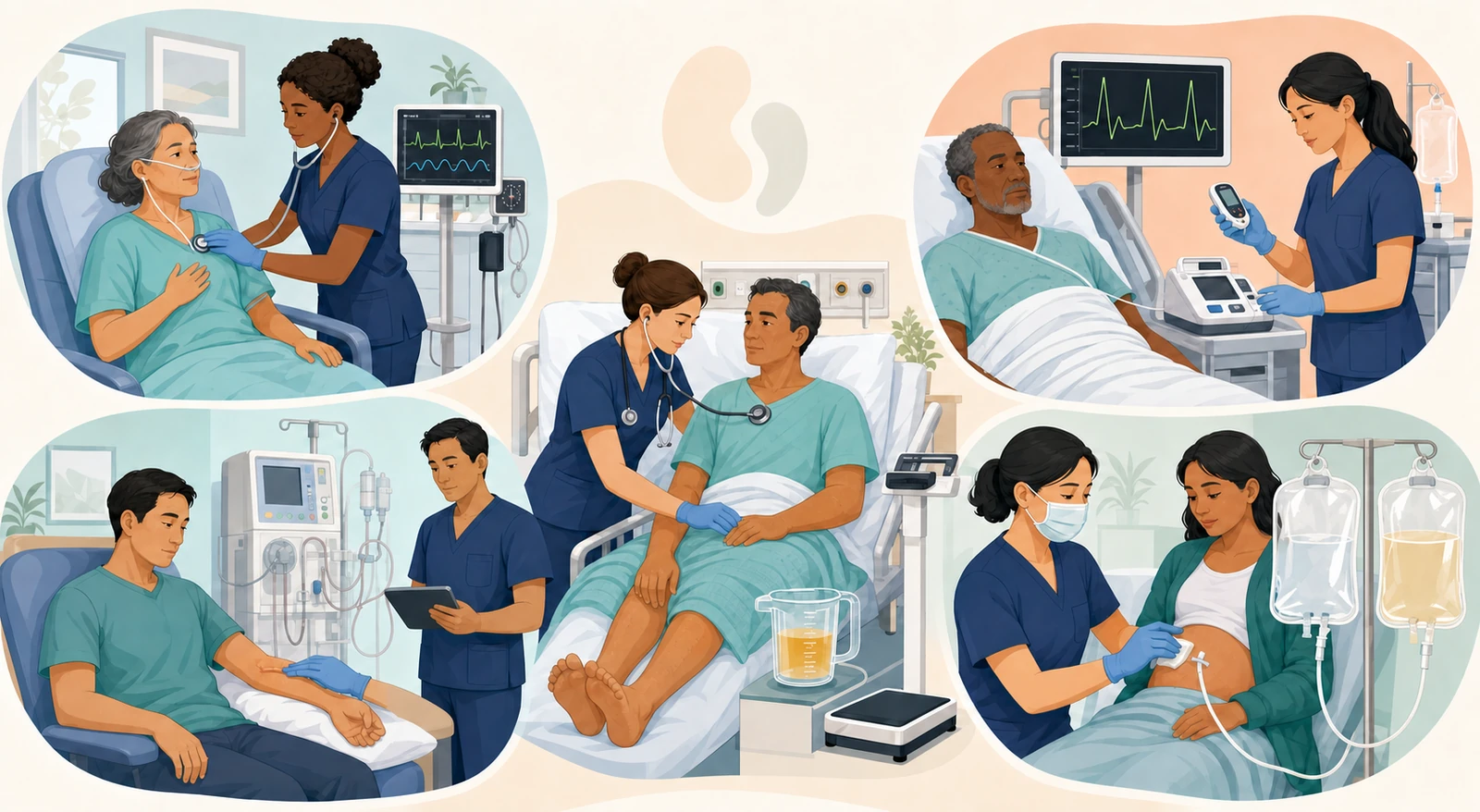
First, check respirations and oxygenation, blood pressure, pulse, skin, and level of consciousness, as well as for shock, sepsis, hemorrhage, and pulmonary edema.
Check hourly urine output and output per body weight, any sudden anuria, catheter patency and bladder distention, and the color, sediment, or presence of blood in the urine.
Differentiate between fluid deficit and overload using daily weight, I/O, edema, crackles, JVD, mucous membranes, and orthostatic changes.
Connect the dots between hypoperfusion, intrinsic kidney damage, or urinary tract obstruction with recent medications, contrast dye, surgery, or infection.
Screen for hyperkalemia, metabolic acidosis, refractory pulmonary edema, and uremic encephalopathy or pericarditis.
Repeatedly compare perfusion, urine output, weight, respiratory status, potassium, acid-base balance, and BUN/creatinine against the prescribed treatment response.
Creatinine may not rise immediately after injury, and not all AKI is oliguric.
The basic KDIGO criteria for recognition are: an increase in creatinine of ≥0.3 mg/dL within 48 hours, an increase to ≥1.5 times baseline within 7 days, or a urine volume of <0.5 mL/kg/h for 6 hours. Even if lab values have only changed slightly, the presence of new-onset oliguria or anuria with decreased perfusion requires an immediate cause evaluation.
| Category | Key Clues | Nursing Connections |
|---|---|---|
| Pre-renal / Hypoperfusion | Hemorrhage, dehydration, sepsis, hypotension, low cardiac output, cool skin, orthostatic changes | Track the response to resuscitation of the underlying cause and perfusion, but reassess fluids if crackles, JVD, or edema develop |
| Intrinsic / Parenchymal Damage | Prolonged AKI, clues of nephrotoxins, sepsis, rhabdomyolysis, or glomerular disease | Review medications, dosages, lab results, and urine findings, and report to stop or adjust nephrotoxic exposures |
| Post-renal / Obstruction | Anuria, intermittent urine stream, bladder distention, BPH, stones, tumors, catheter kinking | Check catheter tubing and position, and verify bladder scan results, imaging, and decompression orders |
| Mixed | Hypoperfusion and parenchymal damage can occur together, such as in sepsis, heart failure, or post-surgery | Don't fixate on a single cause; continuously re-evaluate hemodynamics, fluid status, medications, and infection response |
Oliguria does not automatically equal a fluid bolus.
First, check for catheter kinking, bladder distention, and signs of hypovolemia. When hypoperfusion is suspected and there is no pulmonary edema, administer prescribed isotonic crystalloids and immediately assess the response through blood pressure, peripheral perfusion, urine output, respiratory status, crackles, and JVD. If fluid overload, heart failure, or pulmonary edema is already present, additional fluids can worsen the condition.
Hourly amount, weight-based trend, sudden anuria, hematuria, foaminess, sediment, and catheter patency
Daily weight under the same conditions, accurate I/O, edema, lung sounds, JVD, mucous membranes, orthostatic changes
Potassium, sodium, bicarbonate, BUN/creatinine, glucose, calcium, phosphate, and any prescribed urine studies
NSAIDs, ACEi/ARBs, diuretics, aminoglycosides, contrast dye, supplements, and the time of last administration
Diuretics are not a medication that reverses AKI itself. They may be prescribed for the indication of managing fluid overload, but don't assume kidney function has recovered just because urine output increases. Medication dosages should be reviewed based on current kidney function, dialysis timing, blood pressure, and electrolyte levels.
Consider the possibility of a hemolyzed sample, but if you see muscle weakness, bradycardia, arrhythmias, or ECG changes, don’t delay treatment while waiting for a repeat lab draw. A normal ECG doesn’t completely rule out dangerous hyperkalemia either.
Prepare the prescribed IV calcium right away. Calcium lowers the risk of arrhythmias but doesn’t actually lower the serum potassium level, so the next steps absolutely must follow.
Administer insulin and glucose, and if needed, an inhaled beta-agonist according to your institutional protocol. In renal failure, insulin’s effect can be prolonged, so monitor repeatedly for delayed hypoglycemia.
Depending on renal function and volume status, coordinate diuresis, a potassium binder, or dialysis. The shifting treatments are only temporary, so recheck potassium, ECG, and glucose to watch for rebound.
| Indication axis | Red flags | Nursing actions |
|---|---|---|
| Acidosis | Severe metabolic acidosis unresponsive to medical management | Check ABG/VBG, bicarbonate, respiratory status, hemodynamics; contact the renal and critical care teams |
| Electrolytes | Hyperkalemia or arrhythmias that don’t respond to treatment or keep recurring | Cardiac monitoring, immediate treatment, and prepare for dialysis access, consent, and equipment |
| Fluid | Refractory pulmonary edema or fluid overload with worsening oxygenation | Elevate the head of bed, monitor oxygenation, blood pressure, weight, I/O, and assess for urgent ultrafiltration |
| Uremia | Uremic pericarditis, encephalopathy, bleeding tendency, or severe symptoms | Watch for chest pain, friction rub, altered mental status, seizures, bleeding; prompt specialist team evaluation |
| Toxins | Exposure to a dialyzable toxin with deteriorating systemic condition | Identify the substance, time, and amount; coordinate with poison control and the renal team for detoxification and dialysis route |
Don’t decide to start dialysis based on a single BUN, creatinine, or potassium value alone.
KDIGO and NICE recommend urgently considering renal replacement therapy when there are life-threatening changes in fluid, electrolytes, or acid-base balance. The decision to start is based on the patient’s overall condition, trends over time, and response to treatment.
| Timing | What to check | Changes you can’t afford to miss |
|---|---|---|
| Pre-dialysis | Weight under the same conditions, vital signs and orthostatic changes, lung sounds and edema, potassium and glucose, access thrill/bruit, and skin | Pulmonary edema, hyperkalemia, infection, loss of access blood flow, and medications that need to be adjusted before dialysis |
| During dialysis | Blood pressure, heart rate, level of consciousness, chest pain, dyspnea, cramping, nausea, machine alarms, and access site bleeding | Hypotension, arrhythmias, air or blood loss, severe cramping, fever, and chills |
| Post-dialysis | Weight, vital signs and orthostatic safety, respiratory status and edema, needle site hemostasis, thrill/bruit, and distal perfusion | Persistent hypotension or syncope, dyspnea, uncontrolled bleeding, and new headache, confusion, or seizures |
Look, listen, and feel: check the skin, bruit, thrill, and the color, temperature, and sensation of the distal extremity. If there’s any change or loss of these findings, don’t use the access and report it immediately.
Avoid blood pressure measurements, venipuncture, IV lines, tight clothing, heavy pressure, or sleeping on that arm. Share the access location with a bracelet and during handoff.
After needle removal, apply direct pressure to each site individually, but don’t occlude the entire access flow. If bleeding persists, call for emergency help.
Follow aseptic technique and your institutional protocol when connecting, disconnecting, and changing the dressing at the hub. Immediately report any moisture, contamination, redness, drainage, or fever.
Don’t automatically dismiss post-dialysis fatigue as “normal.”
First, check the blood pressure, orthostatic changes, oxygenation, heart rhythm, access site bleeding, and weight change. Dyspnea, chest pain, syncope, persistent hypotension, or new neurological changes are not just routine fatigue.
Make sure you have a clean, dry space, perform hand hygiene and wear a mask, and use aseptic technique at the connection site. Check the solution’s expiration date, concentration, any leaks or cloudiness, and verify it against the prescription, then warm it to near body temperature using an approved method.
Watch for pain, shortness of breath, leaks, and the inflow rate. Never use a microwave or hot water to heat the solution yourself, and don’t add anything to the solution unless it’s prescribed.
Document the prescribed fill volume, the amount that drained out, how clear it is, any fibrin or blood, and note any abdominal pain, fever, nausea, and the condition of the exit site.
If you see cloudy effluent or new abdominal pain, save the drained fluid to send for cell count with differential, Gram stain, and culture, and report it immediately. Don’t delay treatment while waiting for culture results.
If the drainage is slow: Check for clamps, kinks, the position of the bag, and the tubing, and try changing the patient’s position. Assess for possible constipation, catheter migration, fibrin, or obstruction, but don’t forcefully milk the catheter or irrigate it without a prescription. If new pain, cloudiness, or fever is present, don’t assume it’s just a simple positioning problem.
Don’t “just watch and wait until the cloudy effluent is definite” when it comes to peritonitis.
The ISPD recommends treating cloudy effluent as peritonitis and starting the diagnostic and treatment pathway. A diagnosis is made when at least two of the following are present: effluent WBC count and differential, symptoms, and culture results. After collecting the specimen, promptly connect to your institution’s protocol for empiric antibiotics.
| Focus Area | Assessment | Teaching Principles |
|---|---|---|
| Fluid & Na | Residual urine output, interdialytic weight gain, edema, blood pressure, thirst | Personalize the prescribed limits, teach about hidden fluids and salty foods, and encourage daily record-keeping |
| K | Current K level, ECG and medications, dialysis interval, constipation, and dietary patterns | Align the plan with lab results and a dietitian’s guidance rather than permanently banning a single food |
| Phosphate | Phosphate, Ca, PTH levels, binder prescription and timing of administration | Teach about phosphate additives in processed foods and link binder use directly to meals |
| Protein | Catabolic state in AKI, dialysis modality, wounds, infection, albumin level, and intake | Avoid a blanket low-protein ban, as needs vary by situation, such as protein loss in PD |
The examples below are newly created scenarios for practicing your clinical judgment flow and do not reproduce actual NCLEX questions, answer choices, or correct answers.
First: Reassess oxygenation, blood pressure, perfusion, weight, I/O, and check the bladder and catheter, then report the fluid overload, cardiac function, and AKI progression. The team will re-evaluate the need for additional fluids, diuretics, blood pressure support, and renal replacement therapy.
What you wouldn’t do: Don’t just keep repeating the same fluid bolus simply because the patient is oliguric.
First: Secure cardiac monitoring and emergency help, protect the myocardium with prescribed IV calcium, then concurrently start cellular shift treatments like insulin and glucose along with a K removal pathway. Repeat the K, ECG, and glucose checks.
What you wouldn’t do: Don’t just give calcium and then assume the K level has been lowered.
First: Raise the head of the bed, check oxygenation, cardiac monitoring, K, and ECG, and open the pathway for pulmonary edema resuscitation and urgent dialysis/ultrafiltration.
What you wouldn’t do: Don’t just “wait for the next scheduled dialysis” or finish up with only a routine diuretic.
First: Do not use the access. Check for a bruit, skin condition, pain, swelling, and distal perfusion, and report immediately to the dialysis or vascular team.
What you wouldn’t do: Don’t try to insert a needle to test patency or compress the arm with a blood pressure cuff.
First: Assess vital signs, orthostatic safety, oxygenation, heart rhythm, weight change, and check the access site for bleeding to rule out hypotension, arrhythmia, or residual pulmonary edema.
What you wouldn’t do: Don’t just dismiss it as common post-dialysis fatigue and send the patient home right away.
First: Do not discard the drainage — save it to prepare for cell count, differential, Gram stain, and culture, and report it immediately. Assess vital signs, abdomen, and exit site, and connect to empiric treatment.
Don't: Don't wait until the next exchange or try to resolve it with just fluid intake.
First: Check for tubing kinks, dependent loops, and bag position, and connect to a bladder scan and catheter patency orders. After resolving the obstruction, monitor for sudden large-volume urination and electrolyte changes.
Don't: Don't assume it's renal parenchymal damage and flush the catheter without assessment.
The trap of directly linking one signal to one intervention
Trends in ABCs, perfusion, urine output, fluid status, bladder, medications
ECG, myocardial protection, shifting into cells, removing from body, recheck
Refractory acidosis, electrolytes, pulmonary edema, uremia, toxins
Weight, blood pressure, access, bleeding, hypotension, breathing
Aseptic technique, inflow, dwell, drainage, cloudiness, abdominal pain
Individualized by labs, residual renal function, and dialysis modality
Evidence Scope: Independently written in August 2026 based on the Reduction of Risk Potential and Physiological Adaptation judgment domains of the 2026 NCSBN RN Test Plan, KDIGO AKI and Acute Hyperkalemia reports, NICE AKI guidelines, UK Kidney Association hyperkalemia guidelines, KDOQI vascular access resources, NIDDK dialysis information, ISPD peritonitis guidelines, and CDC dialysis infection prevention recommendations.
NCSBN 2026 RN Test Plan · KDIGO AKI Guideline · NICE Acute Kidney Injury
KDIGO Acute Hyperkalemia Report · UKKA Acute Hyperkalaemia Guideline · KDOQI Vascular Access
NIDDK Hemodialysis · NIDDK Peritoneal Dialysis · ISPD Guidelines · CDC Dialysis Infection Prevention
Content Boundaries: Only recurring study topics were identified from local feedback materials. Actual exam questions, answer choices, correct answers, screens, patient information, source images, and tables were not copied or reproduced. All explanations, cases, judgment sequences, tables, and illustrations were newly written and created.
This material is a summary for nursing exam study and does not replace actual patient diagnosis or treatment orders. In clinical practice, follow the latest lab results, prescriptions, dialysis modalities, vascular access, infection control, hyperkalemia protocols, and the judgment of the renal and critical care specialist team.
다음 이론을 계속 학습하려면 로그인하세요.
로그인하고 계속 학습필기노트, 하이라이터, 메모는 잘 쓰고 있어?
내보내줘운영진이 검토할게요!
마이페이지에서 차단한 회원을 관리할 수 있어요.