Understanding the Hemodynamic Profile
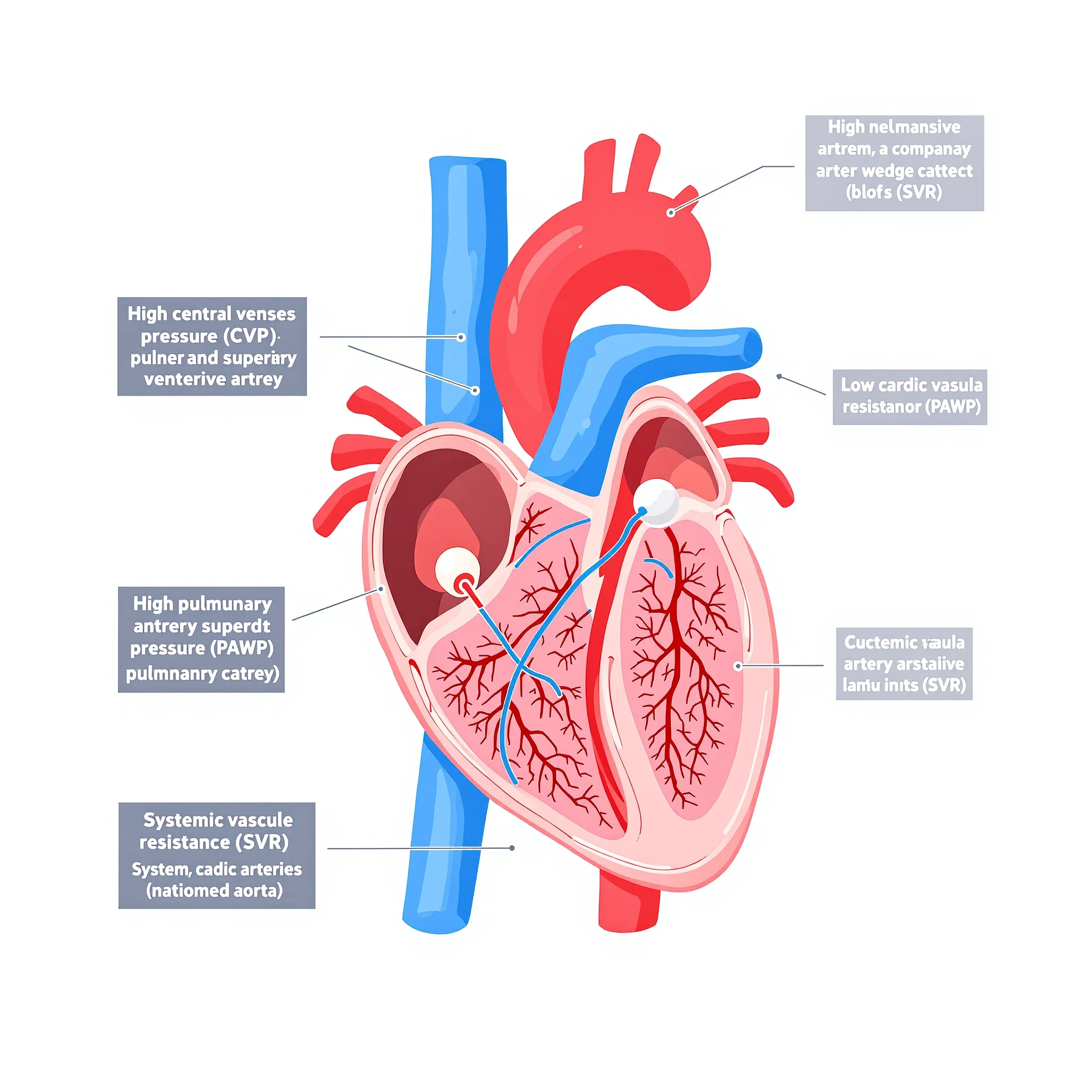
The data presented reflects a classic hemodynamic pattern of cardiogenic shock with significant congestion and poor forward flow. The
central venous pressure (CVP) of
16 mmHg (normal 2–6 mmHg) and
pulmonary artery wedge pressure (PAWP) of
20 mmHg (normal 6–12 mmHg) indicate severely elevated filling pressures, confirming volume overload and pulmonary congestion. Simultaneously, the
cardiac output (CO) is critically low at
3.0 L/min (normal 4–8 L/min), and the
systemic vascular resistance (SVR) is markedly elevated at
1900 dynes/sec/cm⁻⁵ (normal 800–1200 dynes/sec/cm⁻⁵), which represents intense compensatory vasoconstriction [1,2].
Why Increasing IV Fluids (Option 1) is Harmful
Administering a fluid bolus in this scenario directly contradicts the hemodynamic data. The PAWP is already at
20 mmHg, a level at which hydrostatic pressure forces fluid into the pulmonary interstitium, worsening pulmonary edema and respiratory distress. Invasive hemodynamic monitoring in cardiogenic shock is specifically used to phenotype the shock state and avoid such empiric, potentially deleterious interventions
[1]. The elevated CVP and PAWP confirm that the primary problem is not a lack of preload, but rather the failing ventricle's inability to pump the existing volume forward.
Why Reducing Afterload (Option 2) is Not the First Priority
While the SVR of
1900 dynes/sec/cm⁻⁵ represents excessive afterload that further strains the failing left ventricle, vasodilators are not the immediate priority. In the acute "Rescue" phase of cardiogenic shock management, the initial goal is to restore adequate perfusion pressure and cardiac output
[2]. A vasodilator, by rapidly dropping SVR in a patient with a fixed, low stroke volume, can precipitate profound hypotension and coronary hypoperfusion. Afterload reduction is a critical strategy in the later "Optimization" phase once perfusion is stabilized, typically after inotropic or temporary mechanical circulatory support is initiated
[2].
Why Inotropic Support (Option 3) is the Priority
The core defect in this presentation is pump failure, evidenced by the critically low CO of
3.0 L/min despite sky-high filling pressures. The body's compensatory vasoconstriction (high SVR) is an attempt to maintain blood pressure in the face of a failing pump. The priority nursing action is to prepare for inotropic support, such as dobutamine or milrinone, which directly improves myocardial contractility. By augmenting stroke volume, inotropes address the root cause: they increase cardiac output, which in turn promotes forward flow, reduces the compensatory sympathetic surge, and can secondarily lower the elevated SVR and help decongest the lungs by improving renal perfusion [1,3]. This aligns with the "Rescue" phase of the R-O-S-E framework, which focuses on restoring hemodynamic stability
[2].
Why Trendelenburg Position (Option 4) is Contraindicated
Placing a patient with a PAWP of
20 mmHg in the Trendelenburg position is dangerous. This position shifts blood from the lower extremities to the central circulation, further increasing preload and CVP. In a patient already struggling with pulmonary venous congestion, this can acutely worsen respiratory failure by increasing hydrostatic pressure in the pulmonary capillaries. Hemodynamic monitoring data should guide positioning, and in this case, an upright or semi-Fowler's position would be more appropriate to reduce venous return and ease the work of breathing.
References (research sources)
- [1]
Invasive Hemodynamic Monitoring in Acute Heart Failure and Cardiogenic Shock.Research articleBaldetti L, Cosenza M, Galdieri C, Gallone G, Ricchetti G, Gaspardone C, Peveri B, Gramegna M, Cianfanelli L, Calvo F, Pazzanese V, Pieri M, Sacchi S, Ajello S, Scandroglio AM. (2025) · DOI: 10.31083/rcm27034
- [2]
Hemodynamic management of cardiogenic shock in the intensive care unit.Research articleLim HS, González-Costello J, Belohlavek J, Zweck E, Blumer V, Schrage B, Hanff TC. (2024) · DOI: 10.1016/j.healun.2024.03.009