Cardiovascular Medications
ACE Inhibitors & ARBs
- ACE inhibitors (lisinopril, enalapril) block conversion of angiotensin I to angiotensin II, reducing blood pressure and cardiac workload. Monitor for hyperkalemia and angioedema.
- ARBs (losartan, valsartan) block angiotensin II receptors with similar effects but lower risk of cough and angioedema compared to ACE inhibitors.
Memory Aid: "ACE the BP" - ACE inhibitors reduce blood pressure by blocking the conversion process
Beta Blockers
- Metoprolol, atenolol, carvedilol block beta-adrenergic receptors, reducing heart rate, contractility, and blood pressure. Never stop abruptly - risk of rebound hypertension.
- Contraindicated in severe asthma, COPD, and heart block. Monitor for bradycardia and hypotension.
Calcium Channel Blockers
- Amlodipine, nifedipine (dihydropyridines) primarily cause vasodilation. Diltiazem, verapamil (non-dihydropyridines) affect heart rate and contractility.
- Monitor for peripheral edema, constipation, and gingival hyperplasia. Avoid grapefruit juice with certain formulations.
Key Points
- Always check apical pulse before giving cardiac medications
- Monitor electrolytes, especially potassium with ACE inhibitors and diuretics
- Teach patients to change positions slowly to prevent orthostatic hypotension
Heart Failure Management
Medication Therapy
- Diuretics (furosemide) reduce preload by decreasing fluid volume. Monitor for hypokalemia, hyponatremia, and dehydration.
- Digoxin increases contractility and decreases heart rate. Therapeutic level: 0.5-0.9 ng/mL (heart failure); toxicity risk >2.0 ng/mL. Toxicity signs: nausea, visual changes, arrhythmias.
- Aldosterone antagonists (spironolactone) are potassium-sparing diuretics that improve survival in heart failure.
Clinical Scenario: Patient with heart failure on furosemide reports muscle cramps and weakness. Priority action: Check serum potassium level and assess for hypokalemia.
Non-Pharmacological Management
- Sodium restriction (2-3g daily) and fluid restriction (1.5-2L daily) reduce fluid retention and cardiac workload.
- Daily weights at same time with same scale - report weight gain >2-3 lbs in 24 hours or >5 lbs in week.
- Activity modification with gradual increase in exercise tolerance and energy conservation techniques.
Key Points
- Position patients in high Fowler's to reduce preload and improve breathing
- Monitor I&O closely and assess for signs of fluid overload
- Teach patients to recognize early signs of worsening heart failure
Commonly Confused Concepts
| Concept |
Key Difference |
NCLEX Tip |
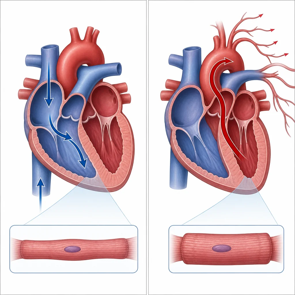
| Preload vs Afterload |
Preload = venous return; Afterload = arterial resistance |
Preload = "before" the heart contracts |
| Systolic vs Diastolic HF |
Systolic = pumping problem; Diastolic = filling problem |
Systolic = "squeeze"; Diastolic = "stretch" |
| Stable vs Unstable Angina |
Stable = predictable; Unstable = unpredictable/worsening |
Unstable = "unpredictable" and urgent |
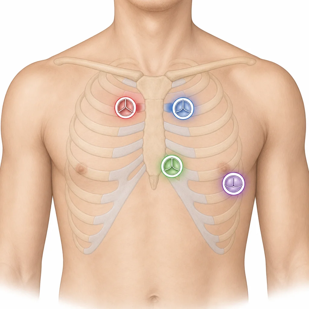
Memory Aid for Heart Sounds:
S1 = "lub" (tricuspid/mitral closure)
S2 = "dub" (aortic/pulmonic closure)
S3 = ventricular gallop (heart failure)
S4 = atrial gallop (hypertension)