컨텐츠 내용을 수정할 수 있습니다
Pediatric emergencies aren't about quickly guessing the diagnosis. They're about spotting time-sensitive clues—like cyanosis, poor perfusion, bilious vomiting, seizures, or altered consciousness—and using them to prioritize stabilization and reporting. Whether it's a cardiac, GI, or neuro issue, your first judgment always starts with the trend in appearance, airway, breathing, circulation, consciousness, and pain.
Core Goal: Make decisions in this order: General appearance & ABC → Perfusion & consciousness → System-specific red flags → Immediate stabilization → Prepare for cause-specific tests/treatment → Repeated reassessment.
Local feedback data only confirmed Kawasaki disease, congenital heart disease, cardiac catheterization, hypertrophic pyloric stenosis, intussusception, Hirschsprung disease, gastric tube care, seizures, febrile convulsions, and increased intracranial pressure as recurring topics. Actual test items, correct answers, answer choices, tables, and images were not reproduced; this content was newly written using current public guidelines from the CDC, AHA, AAP, NIDDK, ASPEN, AES, and Brain Trauma Foundation. Patient-specific oxygen targets, drug dosages, and the timing of tests and surgery will vary depending on age, anatomy, severity, and institutional protocols.
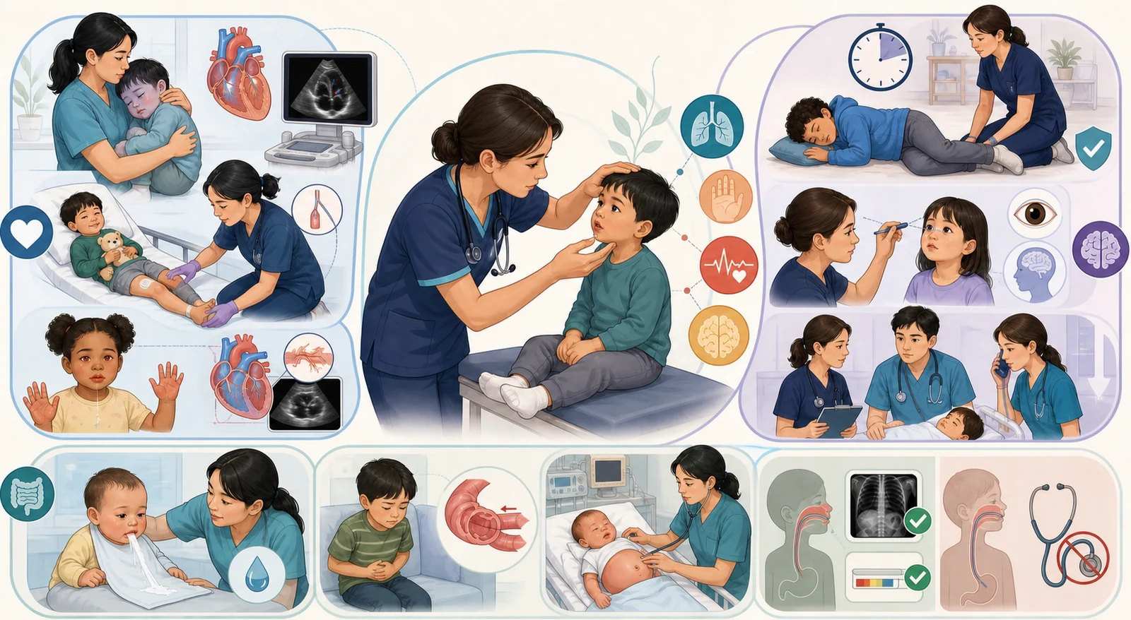
Check if the child makes eye contact with the caregiver, has the strength to speak or cry, responds appropriately to stimuli, or appears limp or abnormally irritable. Distinguish whether a quiet child is stable or has a decreased response due to hypoxia, hypoperfusion, or a postictal state.
Simultaneously assess airway sounds, respiratory rate trends, chest wall movement, retractions, cyanosis, air entry, and oxygen saturation. A single normal pulse oximetry reading does not rule out serious congenital heart disease or ventilation problems.
Connect the dots between heart rate, central and peripheral pulses, skin color and temperature, capillary refill, blood pressure, urine output, hepatomegaly, and edema. Children can compensate with tachycardia and peripheral vasoconstriction before blood pressure drops, so hypotension can be a late sign.
Document baseline status and changes using AVPU or your institution's pediatric neurological tool, and quickly check blood glucose in cases of acute mental status change or seizure. Compare pupils, limb movement, speech, gait, and—for infants—changes in crying and feeding, all adjusted for age.
Check for a rash, abdominal distension, surgical or catheter sites, and signs of trauma to the necessary extent, while keeping the child warm. After any intervention, reassess the same items in the same order to judge if there is actual improvement.
| Observation Cluster | Priority Interpretation | Nursing Action |
|---|---|---|
| Central cyanosis of lips/tongue, low oxygen saturation, clubbing | Possible decreased pulmonary blood flow, right-to-left shunt, or severe hypoxemia | Support airway and breathing, maintain a position of comfort, and activate the lesion-specific emergency pathway |
| Tachypnea, diaphoresis and feeding interruptions, poor weight gain, hepatomegaly | Possible infant heart failure and high metabolic demand | Record the trend of respiratory effort, intake, weight, and urine output rather than focusing on a single feeding volume |
| Pallor/mottling, weak peripheral pulses, cool extremities, decreased urine output | Systemic hypoperfusion or shock | Provide oxygenation, secure vascular access, prepare for glucose and lab tests; administer fluids and medications according to the cause and prescription |
| Pulse or blood pressure difference between upper and lower extremities, weak femoral pulses | Possible left heart outflow tract problem, such as an aortic coarctation lesion | Include four-extremity observation and femoral pulse check and report immediately; do not rule it out based on a passed screening test alone |
When the shunt from the left ventricle to the right ventricle is large, pulmonary blood flow increases, and you may see fast breathing, difficulty feeding, sweating, poor growth, and signs of heart failure. Small defects sometimes close on their own, but the plan for follow-up, medication, or surgery depends on the size and hemodynamics.
A shunt between the atria can cause very subtle symptoms during infancy. Clues include a fixed, widely split S2, exercise intolerance, and frequent respiratory symptoms. You’ll evaluate the defect size and right heart volume load.
A patent ductus arteriosus that shunts from the aorta to the pulmonary artery can cause overcirculation, a wide pulse pressure, bounding pulses, increased work of breathing, and feeding problems. In preterm infants, though, you don’t judge hemodynamic significance by clinical signs alone — you rely on echocardiography.
A large left-to-right shunt that’s been present for a long time can lead to rising pulmonary vascular resistance and shunt reversal. If cyanosis appears later, don’t just brush it off as “part of the original disease.”
After crying, feeding, straining, or dehydration, you may see a sudden worsening of cyanosis along with rapid, deep breathing. The child may become irritable or limp. Don’t wait for the pulse oximetry number — also watch the child’s appearance and level of consciousness.
While a caregiver holds the child or the child is safely supported, bring the knees up toward the chest. This helps raise systemic vascular resistance and reduce the right-to-left shunt. Avoid forcing the child to lie flat or doing anything that makes them cry.
Provide oxygen, continuously monitor the cardiopulmonary status, and call the pediatric cardiac emergency team. Vascular access, fluids, and medications such as sedatives, beta-blockers, or vasoconstrictors — as well as any interventions — depend on the specific lesion, orders, and institutional pathways.
Spells that keep coming back or don’t resolve carry a risk of hypoxic brain injury, so quickly connect with the cardiac team’s plan for a procedure or surgery.
Look at the echo findings together with increasing respiratory support, systemic perfusion, renal function, and gut perfusion. Don’t decide to treat based on a murmur alone.
The current AAP report does not recommend prophylactic or routine early closure within the first 2 weeks of life for preterm infants. If the baby is clinically stable, observation and supportive care may be chosen.
When the PDA is hemodynamically significant, medication for closure is an option. The choice of drug and any contraindications depend on gestational age, kidney and gut status, bleeding risk, and institutional protocols.
If a significant PDA persists beyond 2 weeks, the specialty team individualizes the plan — medication, catheter-based closure, or surgical closure. Intervention is also considered when medical treatment is contraindicated or has failed.
For an infant who uses a lot of energy just to feed, you can plan shorter, more frequent feedings with rest breaks in between. Rather than pushing to finish a set volume each time, watch the respiratory rate, sweating, color changes, and fatigue.
Calorie concentration, time-limited feeds, and tube feedings follow the plan from the cardiac and nutrition teams. Changing formula concentration on your own can cause dehydration or electrolyte problems.
If you mix the medicine into a full feeding bottle and the baby doesn’t finish it, you won’t know how much of the dose was actually given. Use a measuring device and follow the prescription instructions, and never double a missed dose.
Check the apical pulse according to your institution’s policy and the prescriber’s orders. Immediately report bradycardia, rhythm changes, repeated vomiting, or a new feeding intolerance. When toxicity is suspected, evaluate not only the serum level but also potassium, renal function, and any interacting medications.
| Timing | Key Assessments | Red Flags |
|---|---|---|
| Before the procedure | Consent, NPO status, allergies, medications, kidney function, baseline peripheral pulses and skin color | Dehydration, signs of infection, contrast/sedation risks, baseline perfusion asymmetry |
| Immediately after recovery | Airway, breathing, level of consciousness, vital signs, insertion site bleeding/hematoma, distal pulse, color, temperature, capillary refill, sensation, movement | Persistent bleeding, expanding hematoma, a cool and pale limb, weakened pulse, severe pain |
| During bed rest | Limb alignment and activity restrictions according to the access vessel, closure device, and institutional guidelines | Do not apply the same duration of strict bed rest to every child |
| Discharge teaching | Instructions on dressing care, activity, bathing, fluid intake, and who to contact | Bleeding that won't stop, chest pain, shortness of breath, fever, increasing pain or swelling at the site, a cold limb |
| Classic Clue | What to Observe | Interpretation Caution |
|---|---|---|
| Persistent fever | Fever typically lasting 5 days or more | Don't just wait for the full 5 days if characteristic findings and high risk are present |
| Bilateral conjunctival injection | Redness usually without prominent exudate | Distinguish from infectious conjunctivitis where purulent discharge is the main feature |
| Oral changes | Red, cracked lips, strawberry tongue, erythema of the oral mucosa | If pharyngeal exudate or ulcers are the main feature, consider other causes too |
| Rash and extremity changes | Various rashes, erythema and swelling of the hands and feet, periungual peeling during the convalescent phase | Don't wait for skin peeling to make the diagnosis or delay treatment |
| Cervical lymph node | Usually a unilateral, large cervical lymph node | All criteria may not appear at the same time |
If a child has fever and multiple mucocutaneous findings without a typical infectious cause, connect the inflammatory markers, blood and urine findings, and the echocardiogram. Incomplete Kawasaki disease, which doesn't meet all the criteria, can also cause coronary artery complications, so evaluate it using a clinical pathway.
If suspected, connect with the pediatric cardiology team and evaluate the coronary arteries with an echocardiogram. Even if the initial echo is normal, stick to the follow-up plan.
The standard treatment is IVIG and aspirin as directed by a specialist. This is a specialist-treatment exception to the general rule of not casually giving aspirin to children with viral illnesses.
If the coronary artery risk is high or if fever persists or returns after IVIG, the specialist team will individualize treatment with additional IVIG, corticosteroids, or other anti-inflammatory therapies.
After using 2 g/kg of IVIG for Kawasaki disease, vaccines containing measles or varicella components are generally deferred for 11 months. This isn't a rule to defer all live vaccines or all vaccines, and exposure or outbreak situations are adjusted according to the latest immunization guidelines.
| Characteristic | Priority Consideration | First Actions |
|---|---|---|
| Dark green, bilious vomiting | Obstruction below the duodenum, such as malrotation with volvulus—a surgical emergency | Prepare for NPO, gastrointestinal decompression, IV access, tests, and calling pediatric surgery |
| Progressively worsening, projectile, non-bilious vomiting | Hypertrophic pyloric stenosis in early infancy | Assess dehydration, electrolytes, acid-base status, and an ultrasound |
| Intermittent severe pain, pallor, lethargy, with vomiting | Intussusception | Don't wait for bloody stools; prepare for NPO, IV access, imaging, and reduction |
| Distension, fever, foul-smelling diarrhea or explosive stools | Hirschsprung-associated enterocolitis, etc. | Immediate inpatient evaluation, fluids, antibiotics, and a decompression pathway due to sepsis risk |
Usually between 2–8 weeks of age, vomiting after feeding gradually gets worse and turns projectile. Because the obstruction is above where bile enters, the vomit is usually non-bilious, and the baby may act hungry again right after vomiting.
You may see poor weight gain, fewer wet diapers, a sunken fontanelle, and dry mucous membranes. Repeated loss of stomach acid can lead to hypochloremic, hypokalemic metabolic alkalosis.
Ultrasound is the main test. Visible gastric peristalsis or an olive-shaped mass can be clues, but don’t rule it out just because you don’t feel them.
Correct fluid, electrolyte, and acid–base imbalances before pyloromyotomy. After surgery, start and advance feedings according to the surgical plan, and keep a close eye on vomiting, pain, the wound, and hydration status.
You may see sudden severe crying with the legs pulled up toward the belly, quiet periods in between, vomiting, pallor, or unexpected lethargy. A pain-free interval doesn’t mean the problem has gone away.
Stool mixed with blood and mucus, an abdominal mass, and distention can show up, but the classic triad doesn’t always appear. If you wait for bloody stool, bowel ischemia may already be progressing.
Keep the child NPO, secure IV access, and prepare fluids, pain management, and diagnostic studies. If there’s no perforation or peritonitis and the child is stable, image-guided air or contrast enema can serve as both diagnosis and treatment.
If enema reduction fails or there’s perforation, peritonitis, or instability, surgery is needed. Even after successful reduction, watch for recurrence of pain, vomiting, bloody stool, and monitor perfusion.
| Phase | Key point | Safety alert |
|---|---|---|
| Suspicion | Delayed meconium passage, abdominal distention, bilious vomiting, chronic constipation, failure to thrive | Don’t write it off as simple constipation for a long time |
| Testing | Contrast enema and anorectal manometry can provide clues | Rectal biopsy is the definitive test that confirms absence of ganglion cells |
| Definitive treatment | Pull-through surgery to remove the aganglionic bowel and connect normal bowel | Depending on the condition and anatomy, a staged ostomy may be needed |
| Enterocolitis | Fever, rapid distention, foul-smelling explosive diarrhea, lethargy, poor perfusion | Can happen before or after surgery; needs immediate fluids, antibiotics, decompression, and surgical evaluation |
Rectal irrigation is only done using the prescribed method when ordered by the medical team and when the caregiver has been properly trained. Avoid any random enemas, suppositories, or rectal temperature measurements because of the risk of injury and perforation.
Even before feeding, you’ll see excessive drooling, coughing, cyanosis, choking, and a feeding tube that won’t advance. Stop oral feeding, manage secretions from the upper blind pouch, keep the head elevated, prevent aspiration, and prepare for surgery.
In preterm infants, watch for abdominal distention, feeding intolerance, bloody stools, apnea, temperature instability, and poor perfusion. They need NPO, gastric decompression, IV fluids and nutrition, antibiotics, serial abdominal exams and imaging, and close monitoring for perforation.
Don’t handle or try to push the exposed bowel back in. Use a warm, sterile, non-adherent dressing and a protective covering to reduce heat and fluid loss. Maintain temperature and perfusion, decompress the stomach, and get ready to transfer to the surgical team.
Check the appearance of the anus, meconium passage, abdominal distention, and any abnormal fistulas. If you suspect an obstruction, don’t insert a rectal thermometer, suppository, or enema — wait for imaging and surgical evaluation.
Measure and mark the insertion length properly. For a newly inserted tube, imaging confirmation is the most reliable method, based on your facility’s policy and the clinical situation. If the position is unclear, don’t use the tube.
Combine facility-approved methods such as checking the external length and securement, and assessing the pH and appearance of aspirate. After coughing, vomiting, reinsertion, or pulling on the tube, reassess for possible movement.
If you notice a new cough, respiratory distress, drop in oxygen saturation, change in tube length, or inability to aspirate, stop feeding or medication administration right away and recheck.
Look at abdominal distention, vomiting, pain, bowel movements, respiratory status, and systemic perfusion. Don’t automatically stop or push feedings in every child based on a single residual volume number alone.
Clear away dangerous objects nearby and place something soft under the head. Only guide them safely to the floor if they are falling; never forcefully restrain their movements.
Record the start and end times, eye and head direction, which side of the body the seizure started on, any changes in consciousness, breathing, or color, and how they recover.
If possible, turn them onto their side to let secretions drain and loosen anything tight around the neck. Do not put objects, fingers, food, or water into their mouth, and do not force their jaw open.
After the seizure, reassess their airway, breathing, oxygenation, blood glucose, and neurological status. Do not give anything to eat or drink until they are fully awake and can swallow safely.
Start timing the seizure, secure the airway, breathing, and circulation, provide oxygen, attach monitors, and prepare IV or other appropriate access. Check blood glucose. Simultaneously, look for trauma and reversible causes.
Give a benzodiazepine quickly via the route that matches your facility’s protocol and the prescribed order. Be ready for possible respiratory depression, and do not increase repeat doses on your own.
Move on to antiepileptic drugs, further testing, calling the specialist team, and preparing for advanced airway management. Evaluate for underlying causes such as infection, electrolyte or metabolic imbalances, toxicity, trauma, or missed medications.
If the movements have stopped but consciousness does not return as expected, consider ongoing seizure activity, medication effects, hypoxia, or increased intracranial pressure. Connect to further evaluation, such as an EEG.
| Category | Simple Febrile Seizure | Complex, Atypical, or Suspicion of Another Cause |
|---|---|---|
| Patient | A generally healthy child aged 6–60 months with a fever-related seizure | Younger than 6 months or older than 5 years, underlying neurological disorder, or severe systemic illness |
| Features | Generalized, lasts less than 15 minutes, occurs only once in 24 hours, and the child returns to baseline | Focal onset, lasts 15 minutes or longer, recurs within 24 hours, or recovery is not as expected |
| Assessment | Determine the cause of the fever and assess meningitis risk through history and physical exam | Individualize the workup for infectious, metabolic, structural, or toxic causes and additional tests |
| Education | Teach seizure first aid, timing any recurrence, and when to get emergency help | Specialist evaluation and an individualized rescue medication plan |
Antipyretics can ease discomfort but do not reliably prevent febrile seizures from happening again. If you see neck stiffness, a persistent change in consciousness, a non-blanching rash, severe headache or vomiting, or a toxic appearance, do not delay evaluating for meningitis or sepsis just because it is labeled a “febrile seizure.”
Look for unexplained irritability or drowsiness, decreased responsiveness, changes in personality, speech, or gait, headache and vomiting, pupillary changes, and new focal deficits. In infants, clues can include a bulging fontanelle, high-pitched cry, poor feeding, a rapid increase in head circumference, and the “sunsetting” eye sign.
If consciousness progressively declines and you see pupil asymmetry, abnormal posturing, or irregular breathing, consider the risk of herniation. Cushing’s triad—hypertension, bradycardia, and irregular respirations—is a late sign, so do not wait for it to appear.
Maintain the airway, oxygenation, and ventilation, and avoid hypotension, hypoxia, and fever. Once a cervical spine injury is ruled out, keep the head and neck in a neutral position and minimize excessive bending or rotation that could block venous return.
Imaging, hyperosmolar therapy, sedation, ICP monitoring, and surgery all depend on the underlying cause and the specialist team’s plan. Do not use aggressive prophylactic hyperventilation unless it is a short-term rescue measure for impending herniation.
You don't just watch and wait until they settle down. Reduce stimulation and start knee-chest positioning, oxygen, monitoring, and a cardiac emergency call all at the same time.
Don't dismiss this as a normal outcome of bed rest. Immediately reassess the insertion site and distal perfusion, and report it along the vascular complication pathway.
Don't just answer "all live vaccines are off-limits." Check the specific IVIG dose given and the vaccine type, then verify the 11-month interval for measles- and varicella-containing vaccines against current guidelines and with the immunization team.
Rather than rushing straight to surgery, prepare NPO, IV access, correction of dehydration, chloride, potassium, and acid-base balance, plus an ultrasound, then connect to pyloromyotomy.
Don't wait for currant-jelly stool. With intussusception as a possibility, start preparing NPO, IV access, imaging, and air/contrast enema.
See this not as simple worsening constipation but as a risk for enterocolitis and sepsis, and prepare the pathway for admission fluids, antibiotics, rectal washouts, and surgical evaluation.
This does not confirm placement. Hold off on using it, and verify according to facility policy through external length, aspirate pH, clinical status, and imaging if needed.
Don't wait any longer for the seizure to stop on its own. Carry out ABCs, oxygenation, blood glucose check, and the first-line medication pathway for status epilepticus simultaneously, and prepare respiratory support.
This material is a study summary and does not replace individual child diagnosis, prescriptions, oxygen targets, drug dosages, surgical timing, immunization decisions, or institutional protocols. The original sources were used only to confirm the presence of exam topics; questions, answers, answer choices, tables, and images were not reproduced.
다음 이론을 계속 학습하려면 로그인하세요.
로그인하고 계속 학습필기노트, 하이라이터, 메모는 잘 쓰고 있어?
내보내줘운영진이 검토할게요!
마이페이지에서 차단한 회원을 관리할 수 있어요.