컨텐츠 내용을 수정할 수 있습니다
Instead of lumping every new symptom under the single label "chemo side effect," we prioritize protecting airway, neurological, cardiac, and renal function while distinguishing between emergencies caused by the tumor itself and those related to treatment.
Core Objective: We'll follow this sequence for clinical judgment: ABCs & Consciousness → Spinal Cord Compression & SVC → TLS & Hypercalcemia → Infection & Bleeding → Infusion Reaction & Extravasation → Immune Toxicity & Radiation Safety → Re-evaluate Response.
We avoid approaches like "cancer patients always have pain," "just pull the IV if fluid leaks," or "let's wait and see about those abnormal labs after chemo." Instead, we connect the timing of onset, the specific drug given, the tumor's location, vital signs, urine output, and neurological changes as if viewing them all on one screen.
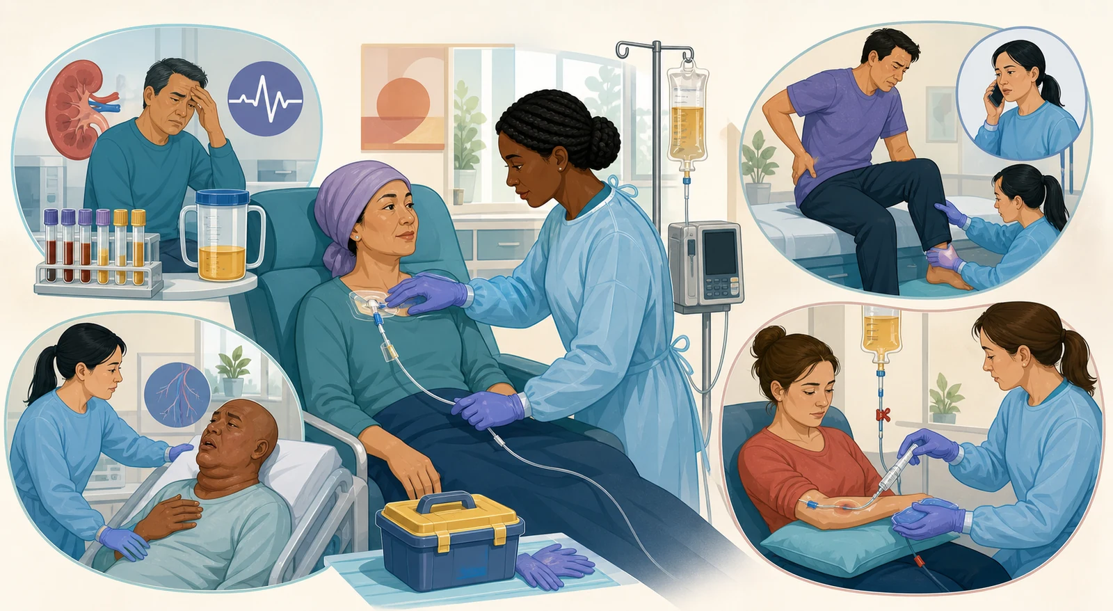
First, check for stridor, dyspnea, hypoxia, chest pain, hypotension, tachycardia, or any new change in consciousness.
Look for locations where compression causes loss of function, such as the spinal cord, airway, great veins, or pericardium.
Connect potassium, phosphate, uric acid, calcium, creatinine, and urine output with ECG and neurological symptoms.
Check for fever, infection, bleeding, anemia, and review the recent trends in CBC, ANC, and platelets.
Confirm when the chemotherapy, immunotherapy, radiation, or surgery started in relation to the symptom, and check the route of administration.
Repeatedly evaluate whether the airway, breathing, circulation, neurological function, urine output, lab values, and infusion site are improving.
Even if a history of cancer seems to explain it, that's not a safe conclusion.
New back pain with leg weakness, facial/neck swelling with dyspnea, decreased urine output with arrhythmia, and pain at the chemo infusion site can each be a time-sensitive problem like spinal cord compression, SVC obstruction, TLS, or extravasation. Don't just give analgesics, oxygen, or antipyretics first and delay the evaluation of the cause.
| Classification | Representative Problem | Functional Threat | First Connection |
|---|---|---|---|
| Structural | Spinal cord compression, SVC syndrome, cardiac tamponade, airway obstruction | Paralysis, respiration, venous return, cardiac output | ABCs, positioning & neuro/cardiopulmonary assessment, urgent imaging & specialist team |
| Metabolic | TLS, hypercalcemia, SIADH | Arrhythmia, seizure, kidney injury, consciousness | ECG, electrolytes & renal function, I/O, prescribed fluids & detoxification strategy |
| Hematologic & Infection | Febrile neutropenia, DIC, bleeding & anemia | Sepsis, shock, organ hemorrhage, hypoxia | Prevent delay in cultures & antimicrobials, assess bleeding, CBC & coagulation |
| Treatment-Related | Infusion reaction, extravasation, immune toxicity, CRS | Anaphylaxis, tissue necrosis, organ inflammation, shock | Decide whether to stop the infusion, ABCs, drug-specific protocol & report to specialist team |
Priority Formula: Before naming the cause, first state the function that could be lost right now. A more complete judgment than simply choosing "potential spinal cord compression" is: "With new weakness and changes in bladder function, there is a risk of permanent neurological injury, so we minimize movement and connect them to an urgent MRI and steroid prescription."
Check for new or progressive, severe localized back pain, pain that worsens with movement or coughing, nighttime pain, and radicular pain.
Document bilateral leg strength, sensation, gait, saddle sensation, reflexes, and any new difficulty with urination or defecation, or incontinence.
If instability is suspected, avoid unnecessary walking, bending, or twisting, and maintain spinal alignment to minimize pain.
Urgently report suspected MSCC and prepare for a whole-spine MRI without delay. If MRI is contraindicated, the specialist team will decide on alternative imaging.
If neurological signs are present, NICE recommends starting dexamethasone as soon as possible. Follow the prescription and institutional protocol for the actual dose and contraindications.
Repeatedly document changes in pain, strength, sensation, gait, and bladder/bowel function before and after the surgical, radiation, or medication plan.
Don’t just give painkillers for back pain in a cancer patient and encourage walking without further assessment.
Especially if new leg weakness, sensory loss, unsteady gait, or changes in urination appear, you need to address the possibility of spinal cord or cauda equina compression first. A simple X-ray alone does not rule out MSCC.
| Clue | What It Means | Nursing Priority |
|---|---|---|
| Swelling of the face, neck, or arms | Decreased venous return from the upper body | Check for bilateral severity, skin color, distended neck/chest wall veins, and any history of IV or central lines |
| Cough, hoarseness, dyspnea, orthopnea | Possible airway or thoracic compression | Keep the patient upright, assess oxygenation, work of breathing, and stridor, and escalate the level of urgency |
| Headache, confusion, syncope, visual changes | Possible cerebral venous congestion or edema | Immediately report changes in consciousness, pupils, or neurological status along with ABCs |
| Worsening when supine | Possible worsening of venous return and airway compression | Maintain a head-up position as tolerated, and avoid unnecessary flat positioning |
Don’t automatically jump to radiation, diuretics, or steroids for every patient with SVC syndrome.
In adults, management depends on the cause, whether there is airway obstruction or cerebral edema, thrombus, tumor histology, and the goals of care. First, assess the ABCs and symptom severity, reduce the burden by keeping the patient head-up, and quickly connect with the oncology, emergency, and interventional teams so they can decide on diagnosis, stenting, anticoagulation, chemotherapy, or radiation.
Watch for muscle weakness, paresthesia, ECG changes, and the risk of fatal arrhythmias.
Can bind with calcium, worsening renal deposition and hypocalcemia symptoms.
Can lead to crystal deposition and acute kidney injury, flank discomfort, and decreased urine output.
Check for tingling, muscle cramps, tetany, seizures, and risk of QT prolongation.
Watch for a vicious cycle where metabolite excretion becomes even harder, and assess fluid tolerance.
This can occur not only after treatment but also before treatment in rapidly growing tumors.
If symptoms and K abnormalities are present, initiate ECG and continuous monitoring, and prepare for arrhythmia and seizure management.
Repeat K, phosphate, Ca, uric acid, creatinine, and LDH based on risk level before and after treatment, and watch the trends rather than a single value.
Administer prescribed IV hydration, maintain strict I/O and daily weights, while simultaneously monitoring for heart failure, renal function, and fluid overload.
Allopurinol or rasburicase is used depending on the risk level and current uric acid. Rasburicase must be avoided in G6PD deficiency.
Manage emergency hyperkalemia treatment and phosphate/calcium levels according to symptoms, ECG, labs, and prescriptions. Do not routinely supplement asymptomatic hypocalcemia.
Do not delay a nephrology and renal replacement evaluation for uncontrolled hyperkalemia, acidosis, fluid overload, symptomatic hypocalcemia, or progressive AKI.
Don’t just keep running fluids or supplements that contain potassium or phosphate uniformly in TLS.
Immediately check lab trends and orders. Also remember, allopurinol is a drug that reduces new uric acid production, which is a different role from rasburicase, which rapidly breaks down existing uric acid.
| System | Clue | Priority Link |
|---|---|---|
| Neurological & Muscular | Fatigue, muscle weakness, drowsiness, confusion, or coma in severe cases | Ensure safety regarding consciousness, falls, and aspiration, and check the rate of change |
| Renal & Fluid | Polyuria, polydipsia, dehydration, changes in urine output, AKI | Monitor I/O, weight, creatinine, and tailor fluids to the cardiopulmonary status |
| Gastrointestinal | Nausea, anorexia, constipation, abdominal pain | Check bowel sounds, intake, and bowel movements, and identify overlapping effects of opioids and dehydration |
| Cardiovascular | Possible shortened QT interval, bradycardia, dysrhythmia | If symptomatic or severe, connect to an ECG and immediate medical orders |
| Bone | Bone pain, pathological fractures | Ensure safety regarding falls and weight-bearing, and assess pain and imaging |
Treatment connection: Since hypovolemia is common, isotonic saline hydration is usually considered first. Calcitonin works quickly but has a short effect, while IV bisphosphonates take longer to act and can be used for sustained control. However, the prescription is individualized based on heart failure status, renal function, albumin or ionized Ca levels, current medications, and the underlying cancer.
Safety checks before administration
Patient education is also part of the pre-administration check. Symptoms to report immediately include burning, pain, or swelling at the infusion site, flushing, itching, throat tightness, dyspnea, chest or back pain, and dizziness. Explain that they should call for help at the very first sign, rather than thinking, “I can just tough it out until it’s done.”
| Category | Key clues | First action | Next decision |
|---|---|---|---|
| Infusion reaction | Flushing, rash, itching, fever, rigors, chest/back pain, dyspnea, wheeze, hypotension | Stop the infusion, assess ABCs and vital signs, call for emergency help and follow the protocol | Determine severity (e.g., anaphylaxis, CRS) and apply drug-specific rescue and restart criteria |
| Extravasation | Site pain, burning, swelling, erythema, blanching, decreased blood return, resistance, or leakage | Stop the infusion, keep the catheter in place, aspirate slowly, do not flush | Determine the drug-specific antidote, apply warm or cold compress, decide on removal timing, specialist referral, and follow-up |
| Infiltration | Non-vesicant fluid, cool and pale swelling, slowing of the infusion | Stop the infusion, assess the site and neurovascular status | Follow the general IV infiltration protocol with elevation and compress |
| Phlebitis | Warmth, redness, tenderness, or a palpable cord along the vein | Stop the infusion and assess whether to remove the catheter | Check for signs of infection or thrombosis and follow the institutional protocol |
As soon as you suspect it, stop the vesicant or irritant infusion. Do not push more fluid through that line or flush it like a normal infusion.
Do not pull it out right away. Slowly aspirate any remaining drug and use the line as a route to administer an antidote if needed.
Check for pain, color, temperature, swelling, blisters, sensation, movement, and distal circulation. Document the length and width, the estimated volume, and the type of access.
Confirm the exact agent, concentration, dose, and whether it is DNA-binding to decide on the antidote and type of compress.
For example, dexrazoxane may be used for anthracyclines, and hyaluronidase for vinca alkaloids. The choice of warm or cold compress follows an agent-specific algorithm.
After the necessary aspiration and antidote, remove the catheter according to the protocol. Report to the provider, pharmacy, and oncology team to plan for photos, education, and long-term follow-up.
The action of “flushing it out with a lot of normal saline” can actually spread the extravasation.
The ONS/ASCO algorithm advises slowly aspirating the remaining fluid and not flushing. A cold compress is used in most cases, but for vinca alkaloids, etoposide, oxaliplatin, and others, a warm compress may be used, so you must identify the drug first.
Chemo gloves, a non-absorbent, closed-front gown, and protective gear specific to the exposure task
Use a biological safety cabinet, CSTD, needleless system, and other measures to reduce exposure
Immediately control the area, use the spill kit and institutional procedures, and manage skin or eye exposure
Handle urine, stool, vomitus, and contaminated linen using hazardous-drug procedures for the designated period
Dispose of tubing, PPE, containers, and sharps in properly labeled waste bins
Provide drug- and institution-specific guidance on home handling, body fluid exposure, and oral medication storage
NIOSH connection: With antineoplastic drugs, occupational exposure can happen not just during preparation and administration, but also when handling contaminated linen, patient body fluids, and waste. Don't rely on a single pair of regular exam gloves for everything—use PPE and engineering controls that match the specific drug, task, and your facility's policy.
| Organ | Warning Clues | Priority Actions |
|---|---|---|
| Lung · pneumonitis | New cough, dyspnea, hypoxemia | Provide oxygenation, rule out infection/PE, and report to oncology immediately |
| GI · colitis | New or worsening diarrhea, abdominal pain, hematochezia or melena | Assess frequency, dehydration, electrolytes, and infection; report without delay |
| Liver · hepatitis | Rising AST/ALT, jaundice, dark urine, nausea | Check lab trends and other causes of liver injury, and connect to whether treatment should be held |
| Endocrine | Severe fatigue, headache, hypotension, weight or temperature changes, abnormal Na or glucose | Evaluate pituitary, thyroid, and adrenal function, and assess for crisis signs |
| Cardiac · Neurologic | Chest pain, arrhythmia, hypotension, new weakness, confusion, seizure | Prioritize ABC, ECG, neuro assessment, and emergency specialist evaluation |
Don't manage immunotherapy-related diarrhea at home the same way you would typical chemo-related nausea and diarrhea.
Immune-related adverse events can appear not only during treatment but also after it ends, and they can affect multiple organs at the same time. While ruling out infection, assess the grade and organ-specific risk, and follow the oncology protocol and orders for immunosuppressive treatment such as steroids.
| Situation | Key Fact | Safety Actions |
|---|---|---|
| External beam | The radiation comes from a machine, so the patient does not become radioactive | No contact isolation needed; focus on skin markings, treatment-site care, and fatigue management |
| Temporary brachytherapy | A sealed source is present while it's inside or near the body | Apply your facility's policies: minimize time, maximize distance, use shielding, and limit visitors |
| Permanent implant | Small sources remain in place, and the radiation decreases over time | Provide discharge teaching based on the nuclide and dose, and follow guidelines for contact with pregnant individuals and infants/young children |
| Source problem or alarm | Possible source dislodgement or retrieval failure | Do not touch or manipulate the source yourself; increase your distance and immediately call the radiation safety team and authorized user |
Never handle a dislodged source with your bare hands or improvised tools.
While keeping the patient safe, reduce your exposure time, maximize your distance, and activate your facility's emergency procedure by calling the radiation safety officer and authorized user. The specific retrieval and shielding equipment, as well as the responsible personnel, depend on the facility license, source type, and written procedures.
Case 1 · New back pain and leg weakness
A patient undergoing treatment for prostate cancer reports back pain that worsens at night, bilateral leg weakness, and new difficulty voiding. This isn't just a request for pain medication—consider possible metastatic spinal cord compression. Minimize movement, establish a neurologic baseline, report urgently, and connect to MRI and dexamethasone orders.
Case 2 · Facial swelling and dyspnea that worsens when lying flat
A patient with a thoracic tumor develops neck and facial swelling, hoarseness, and orthopnea. Suspect SVC obstruction and airway compromise. Prioritize an upright position, oxygenation, and assessment of consciousness and airway, then report to the specialist team immediately.
Case 3 · Decreased urine output and muscle cramps after treatment
A patient with a high tumor burden shows nausea, muscle cramps, and decreased urine output after chemotherapy. Consider possible TLS. Immediately connect to an ECG, check K, phosphate, Ca, uric acid, creatinine, and I/O, and prepare ordered hydration and urate-lowering therapy.
Case 4 · Confusion and polyuria
A patient with bone metastases develops constipation, intense thirst, and polyuria, followed by confusion. Don't chalk this up to opioid side effects alone—suspect hypercalcemia and dehydration. Check mental status, ECG, Ca, albumin/ionized Ca, and renal function.
Case 5 · Burning at the vesicant infusion site
A patient reports burning at the infusion site, and blood return has diminished. Suspect extravasation. Stop the infusion immediately but keep the catheter in place, and gently aspirate. Do not flush. Check the agent-specific antidote and compress algorithm.
Case 6 · Wheezing and hypotension during an infusion
During chemotherapy infusion, flushing, wheezing, and hypotension develop. This is more likely a severe infusion reaction or anaphylaxis than a local site problem. Prioritize stopping the infusion, managing ABC, calling for emergency help, and following the ordered epinephrine protocol.
Case 7 · New cough after immunotherapy
A patient on immune checkpoint inhibitor therapy develops a new dry cough and dyspnea. Along with infection and PE, you should suspect immune-related pneumonitis. Check oxygenation and connect them with the oncology team for evaluation the same day.
Case 8 · Family contact question after radiation therapy
A patient who received external beam radiation asks if it's safe to see their grandchildren. Explain that external beam radiation does not make the patient radioactive. In contrast, brachytherapy and radioactive drugs require separate, source-specific education.
Start with ABCs, consciousness, and location of function loss
Back pain + weakness/sensory/bladder changes → urgent MRI
Face/neck swelling + respiratory distress → upright position, airway
K, P, uric acid ↑, Ca ↓, check ECG and kidneys
STOP → keep catheter in place and aspirate → no flushing
New "-itis" symptoms mean immediate organ assessment
Evidence base: Independently written in August 2026, drawing on the 2026 NCSBN RN Test Plan's Pharmacological and Parenteral Therapies, Reduction of Risk Potential, and Physiological Adaptation categories; NICE 2023 MSCC guidelines; NCI resources on TLS, SVC, and immunotherapy inflammation; the 2025 ONS/ASCO extravasation guideline and 2026 algorithm; and CDC/NIOSH hazardous drug and radiation safety materials.
NCSBN 2026 RN Test Plan · NICE Spinal Metastases and MSCC · NCI Tumor Lysis Syndrome
NCI Superior Vena Cava Syndrome · ONS/ASCO Extravasation Guideline · ONS/ASCO Extravasation Algorithm
CDC/NIOSH Antineoplastic Drug Safety · NCI Immunotherapy Organ Inflammation · ONS Hypercalcemia of Malignancy
NCI External Beam Radiation · CDC ALARA: Time, Distance, Shielding
Content boundaries: Local feedback materials only confirmed recurring study topics like TLS, hypercalcemia, spinal cord compression, SVC syndrome, extravasation, and radiation safety. No actual exam questions, answer choices, correct answers, screens, patient information, source images, or tables were copied or reproduced. All explanations, cases, clinical judgment sequences, tables, and illustrations were newly written and created.
This material is a summary for nursing exam study and does not replace actual patient diagnosis or treatment orders. In clinical practice, follow the latest lab tests, prescriptions, chemo regimens, agent-specific extravasation algorithms, hazardous drug and radiation policies, and the judgment of oncology, emergency, pharmacy, and radiation safety specialist teams.
다음 이론을 계속 학습하려면 로그인하세요.
로그인하고 계속 학습필기노트, 하이라이터, 메모는 잘 쓰고 있어?
내보내줘운영진이 검토할게요!
마이페이지에서 차단한 회원을 관리할 수 있어요.