컨텐츠 내용을 수정할 수 있습니다
Rather than jumping to name the exact cause of abdominal pain, first rule out bleeding, perforation, obstruction, sepsis, and organ dysfunction. Then, connect variceal bleeding, ascites, hepatic encephalopathy, pancreatitis, cholangitis, and ostomy care into one unified priority framework.
Core Goal: Think in this order: ABCs & Hemodynamics → Bleeding, Perforation, Obstruction, Sepsis → Pain Location & Associated Signs → Diagnostics → Endoscopy, Decompression, Drainage → Nutrition & Medications → Reassessment of Response.
Don't jump to conclusions based on pain intensity alone, a single blood pressure reading, or one ammonia level. Look at the whole picture: level of consciousness, skin perfusion, urine output, respiratory status, abdominal findings, estimated blood loss, drainage, and changes over time.
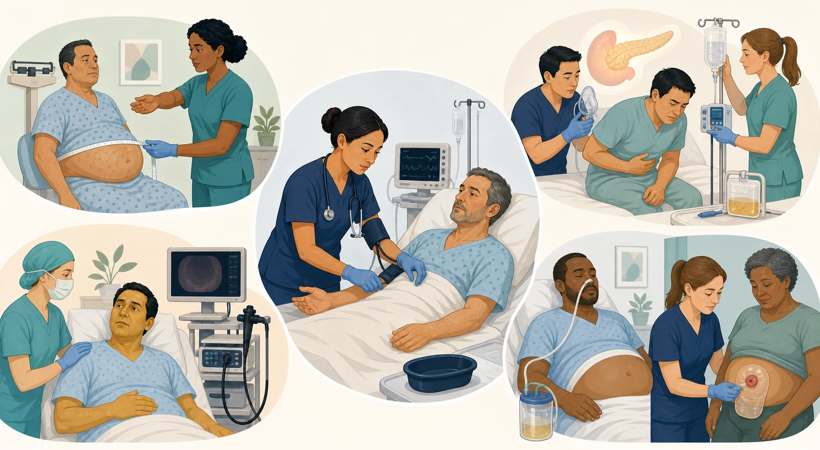
Check airway patency, breathing and oxygenation, pulse, blood pressure, skin signs, level of consciousness, urine output, and any signs of active bleeding.
Screen for massive hemorrhage, peritonitis/perforation, complete obstruction/ischemia, cholangitis/sepsis, and acute hepatic or pancreatic organ failure.
Cluster the findings: epigastric, right upper quadrant, or lower abdominal pain; radiation to the back; relationship to meals; vomiting, stool, flatus, jaundice, and fever.
Connect the need for CBC, coagulation studies, electrolytes, renal and hepatic function, lipase, blood type and crossmatch, imaging, and endoscopy.
Prepare for hemostatic endoscopy, variceal ligation, GI decompression, biliary drainage, or surgical/interventional radiology pathways.
Repeatedly compare level of consciousness, pain, abdominal findings, bleeding, drainage, urine output, BUN/hematocrit, electrolytes, and response to treatment.
A single normal blood pressure reading does not guarantee stability.
In early hemorrhage, compensatory mechanisms can maintain blood pressure. Watch for syncope, dizziness, tachycardia, cool and clammy skin, confusion, narrowing pulse pressure, and decreased urine output. Combine these with trending vital signs so you don't miss hidden shock.
| Clue | Interpretation | Priority Connection |
|---|---|---|
| Hematemesis / Coffee-ground emesis | Suggests active or recent upper GI bleeding | Airway protection, aspiration risk, hemodynamics and bleeding trends |
| Melena | Likely digested blood; can persist for some time even after bleeding stops | Don't estimate current bleeding rate by stool color alone; assess systemic perfusion |
| Bright red hematochezia | Commonly lower GI bleeding, but possible with rapid, massive upper GI bleeding | Prioritize shock and active bleeding assessment over guessing the location |
| Syncope, tachycardia, decreased urine output | Possible circulating volume depletion and organ hypoperfusion | Large-bore IV access, labs and crossmatch, prescribed fluids/blood, and endoscopy prep |
| Anticoagulants / Liver disease | Increased risk for ongoing bleeding, varices, and coagulopathy | Time and dose of last intake, INR, platelets, liver function, and report to provider |
Massive hematemesis and decreased consciousness raise the risk of aspiration and airway loss. Secure oxygenation, suction setup, monitoring, and large-bore IV access. Track serial vital signs, mental status, and urine output.
Prepare CBC, coagulation panel, electrolytes, BUN/creatinine, liver function tests, and type and crossmatch. Confirm any history of anticoagulants, NSAIDs, alcohol use, or liver disease.
For unstable, severe upper GI bleeding, endoscopy is performed immediately after resuscitation. For other upper GI bleeds, endoscopy is generally recommended within 24 hours. Don't put off resuscitation or preparation for definitive hemostasis.
Initial hemoglobin may not immediately reflect actual blood loss. Judge the response by estimated blood loss, mental status, skin signs, urine output, hemodynamics, and serial lab trends.
Stabilize ABCs and hemodynamics, and immediately assess the need for airway protection if there's massive hematemesis or decreased consciousness.
From the moment you suspect it, quickly prepare prophylactic antibiotics and vasoactive therapy according to your institution's protocol.
Prepare for urgent endoscopy and esophageal variceal band ligation. Follow the prescribed orders and protocol for fluid administration and transfusion goals.
If hemostasis fails or rebleeding occurs, a specialized team will evaluate whether rescue therapies like TIPS are appropriate.
Balloon tamponade is not a routine first-line choice.
It's a temporary bridge to buy time until definitive treatment in critical situations where endoscopic hemostasis has failed or is immediately unavailable. It is never initiated without airway security, a skilled team, and intensive monitoring, and you must watch for risks of aspiration, airway obstruction, and tissue injury.
| Assessment | Standardized conditions | Signs of worsening |
|---|---|---|
| Weight | Same time, clothing, and scale; after voiding if possible | Rapid increase, or excessive loss with diuresis causing dizziness or worsening renal function |
| Abdominal girth | Same position, phase of respiration, and anatomical landmark | Increasing distension, pain, dyspnea, or early satiety |
| Respiratory & perfusion | Oxygenation, respiratory rate, peripheral edema, I/O, blood pressure | Hypoxemia, hypotension, decreased urine output, new decline in renal function |
| Infection | Temperature, abdominal pain/tenderness, mental status, WBC with cultures, ascitic fluid studies | New abdominal pain, confusion, or AKI can signal SBP even without fever |
Nursing care for ascites: Monitor the prescribed sodium restriction and spironolactone-based diuretic regimen, adding a loop diuretic if needed. Track weight, I/O, blood pressure, Na/K, and creatinine. For tense ascites, prepare for whether large-volume paracentesis and albumin are ordered. Fluid restriction is not automatically applied to all ascites patients; check for individual indications like hyponatremia.
Check airway, oxygenation, and blood glucose, and rule out other causes like stroke, medications, infection, hypoxia, or electrolyte imbalances. Compare confusion, sleep inversion, asterixis, and behavioral changes to the patient's baseline.
Look for GI bleeding, infection, constipation, dehydration or excessive diuresis, electrolyte shifts, sedatives/opioids, and medication non-adherence.
Aim for the prescribed number of soft stools, documenting mental status and bowel movements together. Severe diarrhea, hypovolemia, hyponatremia, or hypokalemia can actually promote recurrence, so adjust immediately.
For persistent or recurrent cases, check if rifaximin is added, and have the patient and caregiver explain back the precipitating factors, bowel movement goals, signs of dehydration, and medication regimen.
| Monitoring focus | Dangerous changes | Nursing connection |
|---|---|---|
| Circulation | Hypotension, tachycardia, decreased urine output, rising BUN/hematocrit | Administer prescribed balanced crystalloids, avoiding both under- and over-resuscitation, and reassess frequently |
| Respiration | Tachypnea, hypoxemia, dyspnea, new crackles | Evaluate oxygenation and fluid response together, assessing for organ failure and fluid overload |
| Pain & abdomen | Severe epigastric pain, radiation to back, peritoneal signs, persistent vomiting | Manage analgesia and nausea, while differentiating perforation, ischemia, or biliary etiology |
| Metabolic | Signs of hypocalcemia, hyperglycemia, electrolyte abnormalities | Monitor ECG, neuromuscular symptoms, blood glucose, electrolytes, and prescribed replacements |
| Biliary & infection | Jaundice, fever, sepsis, persistent biliary obstruction | If cholangitis is present, prepare for antibiotics and urgent ERCP/drainage pathway |
Correct hypovolemia, but during the first 6–12 hours, reassess vital signs, BUN, hematocrit, urine output, and respiratory status repeatedly to adjust the required volume.
In mild cases, if nausea and pain allow, you can start a low-fat solid diet early, within 24–48 hours. Keeping the patient strictly NPO for a prolonged period until the pain completely disappears is not the standard approach.
Do not routinely use prophylactic antibiotics for sterile pancreatitis without evidence of infection. Check for specific indications like cholangitis or infected necrosis.
Do not automatically perform ERCP on every patient with gallstone pancreatitis. If there is concurrent cholangitis or persistent biliary obstruction, assess the need for urgent decompression.
Organ function takes priority over an elevated lipase level.
Hypotension, hypoxia, decreased urine output, altered mental status, and worsening BUN/hematocrit are signals for immediate reassessment. Also, avoid a fixed formula of rapidly infusing large volumes of fluid; instead, consider the patient's age, cardiac and renal status, and signs of fluid overload.
| Condition | Key Clues | Priority |
|---|---|---|
| Cholecystitis | RUQ pain/tenderness, worsens after eating, nausea, fever | Assess pain, infection, and fluid status; ultrasound and surgical pathway |
| Cholangitis | RUQ pain, fever, jaundice; may also have hypotension and confusion | Sepsis resuscitation, antibiotics, early biliary decompression |
| Biliary Obstruction | Jaundice, dark urine, pale stools, pruritus, cholestatic lab pattern | Identify the location and cause of the obstruction and check for concurrent infection |
| Gallstone Pancreatitis | Epigastric/back pain, elevated lipase, biliary clues | Assess pancreatitis-related organ failure and differentiate from cholangitis or persistent obstruction |
Don't wait for the full Charcot triad to appear.
Elderly or immunocompromised patients may not show a typical fever or severe pain. If you see jaundice or a cholestatic lab pattern, new confusion, hypotension, signs of infection, and biliary dilation together, quickly connect this to sepsis and the need for source control. The ASGE considers cholangitis an emergency and recommends endoscopic biliary decompression within 48 hours if possible.
| Clue | Concern | First Action |
|---|---|---|
| Distention, vomiting, cessation of gas/stool | Mechanical obstruction or ileus | NPO, IV fluids/electrolytes, prescribed NG decompression, imaging/surgical evaluation |
| Pain changing from colicky to constant | Possible strangulation or ischemia | Monitor pain trend, lactate, perfusion, peritoneal signs, and report for urgent surgery |
| Board-like abdomen, rebound tenderness | Perforation, peritonitis | ABCs, sepsis pathway, NPO, IV, antibiotics, prepare for emergency surgery |
| Fever, tachycardia, hypotension, confusion | Sepsis and decreased organ perfusion | Obtain cultures, administer antibiotics, and manage fluid responsiveness alongside source control |
Remember the contraindications: If obstruction or perforation is suspected, do not assume it's simple constipation and give laxatives or an enema, nor apply heat to the abdomen. For an NG tube, verify the prescribed position, drainage, and suction settings, check the oral/nasal skin and I/O, and do not arbitrarily clamp or irrigate it.
Pink or red, moist, and slightly shiny. Initial edema and a small amount of bleeding when wiping can occur, but document the trend.
A stoma that is pale, gray, blue, purple, or black, or one that feels cold or is progressively darkening, requires assessment for possible ischemia.
Keep the peristomal skin dry and intact. Cut the opening just slightly larger than the stoma, and correct any leakage, pain, or rash in collaboration with a WOC nurse.
High-volume, watery output from an ileostomy increases the risk of dehydration and Na/K loss. Monitor the amount, consistency, urine output, thirst, dizziness, and weight.
When there's no output, don't just look at the stoma itself.
If cramping, abdominal pain, nausea, and vomiting occur along with a cessation of gas and stool, assess for a possible obstruction. Also, immediately notify the specialist team for acute retraction or prolapse of the stoma, persistent heavy bleeding, or severe watery output with dehydration. Do not perform routine enemas through an ileostomy.
The example below is a newly created situation for practicing clinical judgment flow and does not reproduce actual NCLEX questions, answer choices, or correct answers.
First: Suspect compensated hemorrhage. Check the airway, perfusion, repeat vital signs, level of consciousness, and urine output. Establish a large-bore IV line, and open the pathway for CBC, coagulation studies, type and crossmatch, and endoscopy.
What not to do: Do not classify the patient as stable based on a single normal blood pressure reading.
Do this first: Check airway, blood glucose, and neurological emergencies. Look for and treat triggers like bleeding, infection, constipation, dehydration, electrolyte imbalances, and medications. Track lactulose response and bowel movements.
Don't do this: Don't just wait for ammonia levels or automatically restrict all protein.
Do this first: Confirm orders and tolerance, then start early low-fat oral intake. Reassess pain, nausea, abdominal status, blood glucose, and fluid balance.
Don't do this: Don't automatically keep the patient NPO long-term just waiting for lipase to normalize.
Do this first: Prioritize organ failure and hypoperfusion. Immediately reassess oxygenation, hemodynamics, BUN/hematocrit, urine output, and fluid response. Prepare for a higher level of care.
Don't do this: Don't check just the pain score or amylase changes first.
Do this first: Treat this as sepsis from cholangitis. Start resuscitation, get cultures, give antibiotics, and prepare for urgent biliary decompression—all at the same time.
Don't do this: Don't delay source control while waiting for a full fever spike or confirmed lab results.
Do this first: Assess for obstruction, ileus, or ischemia. Keep the patient NPO, give IV fluids and electrolytes, and prepare for prescribed NG decompression, imaging, and a possible surgical pathway.
Don't do this: Don't assume it's simple constipation and give a laxative or enema first.
Do this first: Immediately assess stoma perfusion and check for obstruction. Report to the surgical team and WOC nurse. Communicate the color, temperature, swelling, output, pain, and time of onset.
Don't do this: Don't dismiss it as normal postoperative swelling or insert anything into the stoma.
The trap of applying one number or memorized intervention to every patient
Mental status, skin, pulses, blood pressure, urine output, bleeding trends
Constant pain, rigidity, rebound tenderness, fever, sepsis
Bleeding, infection, constipation, dehydration, medications, and lactulose response
Organ failure, fluid response, early nutrition, indications of infection
Jaundice, infection, obstruction along with antibiotics and early decompression
Flatus, stool, vomiting, decompression; color, perfusion, output, skin
Evidence base: Independently written in August 2026, guided by the Management of Care, Reduction of Risk Potential, and Physiological Adaptation judgment domains of the 2026 NCSBN RN Test Plan, the ACG Acute Pancreatitis Guideline, AASLD cirrhosis and hepatic encephalopathy education, NICE upper GI bleeding guidelines, ASGE cholangitis guidelines, and NIDDK guidance on intestinal obstruction and ostomy complications.
NCSBN 2026 RN Test Plan · ACG Acute Pancreatitis Guideline Highlights · AASLD Outpatient Cirrhosis · AASLD Hepatic Encephalopathy
NICE Acute Upper GI Bleeding · NICE Variceal Bleeding Bridge Therapy · ASGE Cholangitis Guideline
NIDDK Intestinal Obstruction · NIDDK Ostomy Complications · MedlinePlus Ileostomy Stoma Care
Content Boundaries: In the local feedback materials, only the recurring study topics were identified. No actual exam questions, answer choices, correct answers, screens, patient information, source images, or tables were copied or restored. All explanations, case examples, judgment sequences, tables, and illustrations were newly written and created.
This material is a summary for nursing exam study and does not replace actual patient diagnosis or treatment orders. In clinical practice, follow the latest prescriptions, test results, your institution's protocols for bleeding, sepsis, liver disease, pancreatitis, surgery, and ostomy care, and the judgment of the responsible specialist team.
다음 이론을 계속 학습하려면 로그인하세요.
로그인하고 계속 학습필기노트, 하이라이터, 메모는 잘 쓰고 있어?
내보내줘운영진이 검토할게요!
마이페이지에서 차단한 회원을 관리할 수 있어요.