컨텐츠 내용을 수정할 수 있습니다
Before you even think about the diagnosis, check for bleeding and distal neurovascular status first. Then, rule out limb-threatening conditions like compartment syndrome and open fractures. After that, you'll connect the dots through immobilization, traction, post-op mobility, and amputation rehab.
Core Goal: Think in this order: Systemic stability → Distal neurovascular trends → Compartment syndrome & open fractures → Immobilization device safety → Thrombosis & infection → Prescribed mobility and rehab.
Don't just stop at "the pain is severe" or "they had surgery." You need to repeatedly compare the location, quality, and trend of the pain, its relationship to passive stretch, changes in sensation, motor function, color, and temperature, as well as device alignment and weight-bearing orders—all under the same conditions.
Check the airway, breathing, circulation, active bleeding, signs of shock, and associated injuries first.
Compare color, temperature, capillary refill, pulses, sensation, and motor function on both sides, and document them along with the time.
Pay separate attention to pain that's out of proportion to the injury, pain that increases despite analgesia, and pain with passive stretch.
Immediately screen for compartment syndrome, open fractures, vascular or nerve injury, excessive swelling, and external compression.
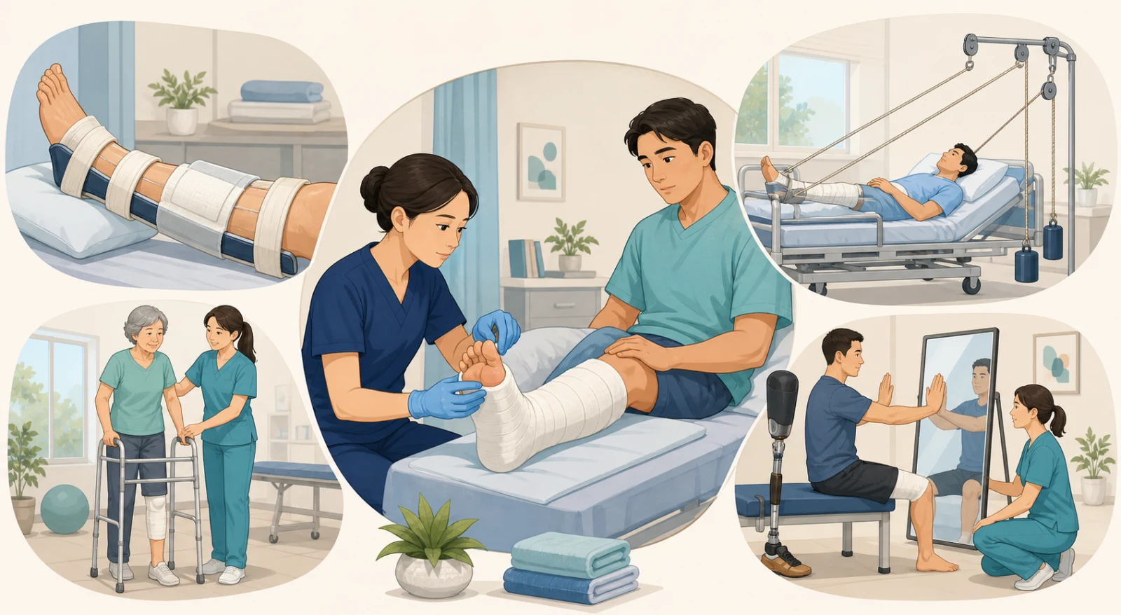
Check the alignment, pressure points, and skin integrity for splints, casts, and traction, along with the ropes, pulleys, weights, and prescribed weight-bearing status.
Prevent thrombosis, infection, pressure injuries, and contractures, and begin pain management, mobility, and exercise as prescribed.
A palpable pulse does not rule out compartment syndrome.
Pain that's out of proportion and steadily increasing, pain that worsens when you passively extend the fingers or toes, and new-onset tingling or loss of sensation can be earlier warning signs. Pallor, paralysis, and pulselessness may appear only with advanced ischemia, so don't wait for all the signs to gather.
| Assessment Item | What to Look For Specifically | Dangerous Changes |
|---|---|---|
| Pain | Location, intensity, quality, onset, response to analgesia and immobilization, and its relationship to passive stretch | Pain disproportionate to the injury or steadily increasing; worsening despite pain medication |
| Sensation | Tingling, burning, numbness; compare light touch on the same spot on both sides | New sensory changes or a sensory deficit that is expanding in area |
| Motor | Active movement of fingers/toes; compare strength and pain to the opposite side | New weakness, inability to move, or pain that sharply increases with movement |
| Perfusion | Color, temperature, capillary refill; compare palpable or Doppler pulses at the same point | Cool, pale, or bluish color; delayed refill; a previously present pulse becoming weaker or absent |
| Device & Edema | Cast edges, tightness of dressings/splints, extent of swelling, skin pressure points | Rapidly increasing swelling, localized burning pain, suspected pressure or drainage under the device |
If you see excessive, increasing pain, pain with passive stretch, a firm, tense compartment, or new sensory/motor changes, treat it as an emergency.
Activate the orthopedic or emergency response pathway and accurately communicate the time the changes started and your serial assessment findings. The longer the delay in diagnosis and decompression, the greater the risk of tissue damage.
If you suspect a constrictive dressing or cast, don't remove the device on your own. Notify the provider immediately, and then prepare for splitting or loosening according to your facility's emergency protocol.
Avoid excessive elevation, which can further compromise arterial perfusion, and generally keep the limb at heart level. Follow orders regarding NPO status, IV access, and surgical prep, keeping the possibility of an emergency fasciotomy in mind.
Don’t just give another painkiller and wait to see if it works.
Pain relief is necessary, but it shouldn’t become a waiting strategy that masks the underlying cause. Immediately report any increase in pain and pain with passive extension along with new neurovascular changes, and keep reassessing.
Control bleeding and cover the wound with a sterile dressing. Don’t push protruding bone back in or irrigate the wound deeply at the scene. Link infection risk, tetanus, and the surgical pathway.
Immobilize the injury as prescribed and compare neurovascular status before and after. Keep them dry, and never put anything inside—no objects, no powder.
Check cast edges, heels, and the sacrum. Report any localized burning sensation, foul odor or drainage, hot spots, numbness, pallor, coolness, or decreased movement.
Maintain body alignment and countertraction. Keep the rope in the pulley groove and the weight hanging freely—never resting on the floor or bed. Don’t lift or remove the weight on your own.
Traction works only when it’s continuous. If a knot catches on the pulley, the weight touches the floor, or the patient slides down the bed, the prescribed pull is lost. First, check the patient’s alignment, breathing, skin, and neurovascular status. Then restore the equipment pathway and report any abnormalities. For skeletal traction, also check the pin site and signs of infection according to your facility’s guidelines.
| Situation | Nursing focus | Don’t apply the same rule to everyone |
|---|---|---|
| Hip fracture surgery | Pain control, delirium/pressure injury/VTE prevention, verify weight-bearing orders, PT evaluation and mobilization by the next day if no contraindications | Extending bed rest just because the patient is older |
| Hip replacement | Confirm the surgical approach, prescribed movement restrictions and duration, weight-bearing status, and assistive device at every handoff | Automatically applying the same 90° flexion, internal rotation, and adduction restrictions to every patient |
| Knee replacement | Prescribed ROM, ankle pumps and quadriceps sets, ambulation, monitor pain, swelling, wound, and VTE | Keeping a pillow under the knee for long periods, which holds it in constant flexion |
| Shoulder surgery | Sling/abduction pillow positioning, neurovascular status of the hand, prescribed hand, wrist, and elbow exercises | Starting shoulder ROM, pulling, or lifting without checking the specific procedure |
“Hip surgery = always the same precautions” is not a safe formula.
The AAOS explains that the necessary restrictions and their duration vary with the surgical technique, and not every surgeon recommends the same precautions. Confirm the procedure name, approach, weight-bearing status, assistive device, and individual orders. Then have the patient explain and demonstrate their own restrictions.
| Type | What it means | Nursing connection |
|---|---|---|
| Immediate post-op pain | Acute pain from the incision and tissue injury | Assess pain, bleeding, swelling, infection, and response to analgesia |
| Residual limb pain | Pain felt in the remaining part of the limb | Check the wound, skin, swelling, pressure, infection, and prosthetic fit together |
| Phantom limb pain | Real pain felt in the amputated part | Don’t dismiss it—assess the pattern and combine medications, non-drug measures, and rehab interventions |
| Phantom limb sensation | A non-painful feeling that the amputated limb is still there | Explain that it’s a common experience and teach fall and mobility safety |
| Related musculoskeletal pain | Back or contralateral limb pain from compensatory movements and overuse | Re-evaluate gait, posture, strength, and prosthetic use with the rehab team |
Follow orders and facility protocols for dressing, compression, and shaping. Check skin color, temperature, drainage, the wound, and distal compression every time.
Plan alignment and ROM so the hip and knee aren’t kept in prolonged flexion. The timing of prone positioning or exercises is individualized based on the surgery, wound, comorbidities, and rehab team orders.
Don’t rely on medications alone. Combine compression, exercise, desensitization, mirror or graded motor imagery, sleep, and emotional support with the team.
Ask first if the patient is ready to look at the residual limb, and give them choices. Address grief, anxiety, body image, functional goals, and family/peer support together.
The examples below are newly created scenarios for practicing clinical judgment flow and do not reproduce actual NCLEX questions, answer choices, or correct answers.
First: Suspect compartment syndrome, report it immediately as an emergency, and communicate the bilateral neurovascular status along with the time of any changes. Avoid elevating the limb excessively, and prepare for pressure relief and the surgical pathway.
What not to do: Do not feel reassured just because a pulse is present, and do not delay reassessment while waiting for analgesic effects.
First: Check the patient’s alignment and the skin and distal neurovascular status, and safely restore the bed, line, pulley, and weight path so that the prescribed traction remains continuous.
What not to do: Do not arbitrarily remove the weight or rest it on the bed.
First: Verify the surgical approach, the surgeon’s written restrictions and their duration, and the weight-bearing orders. Consolidate them into one unified plan and share it with the patient and the team.
What not to do: Do not automatically apply the familiar posterior-approach restrictions to every patient.
First: Check the orders to avoid sustained knee flexion, and support the heel if needed to promote extension. Carry out ankle pumps, quadriceps sets, ROM, and ambulation according to the PT plan.
Reason: Staying in a flexed position solely because of pain can interfere with knee extension recovery and function.
First: Acknowledge phantom limb pain as real pain and assess its intensity, quality, and impact on function. Separately check the residual limb’s condition and connect the patient to prescribed pharmacologic, non-pharmacologic, and rehabilitation interventions.
What not to do: Do not dismiss it as confusion or exaggeration.
First: Reassess the pain, confirm pre-mobilization analgesia, orthostatic changes, and weight-bearing orders, and then begin safe mobilization using PT and assistive devices.
Reason: Unless there is a medical or surgical contraindication, rehabilitation and daily mobilization by the day after surgery are recommended.
The trap of applying one memorized sentence to every patient
ABCs, bleeding, shock, and associated injuries
Pain, passive extension, sensation, motor function, perfusion trends
Compartment syndrome, open fracture, vascular injury
Alignment, pressure points, line, pulley, freely hanging weight
Approach, weight-bearing, individualized restrictions, early mobilization
Acknowledge pain, residual limb, contractures, VTE, infection, functional goals
Evidence Scope: Independently written in August 2026 based on the Safety and Infection Prevention and Control, Reduction of Risk Potential, and Physiological Adaptation judgment areas of the 2026 NCSBN RN Test Plan; AAOS guidance on fractures, casting, and hip and knee surgery; the AHRQ patient safety review on acute compartment syndrome; NICE recommendations for mobilization after hip fracture; and the VA/DoD rehabilitation guideline for lower limb amputation.
NCSBN 2026 RN Test Plan · AAOS Adult Forearm Fractures · AHRQ Acute Compartment Syndrome
AAOS Activities After Hip Replacement · AAOS Total Knee Replacement Exercise Guide · NICE Hip Fracture Recommendations · VA/DoD Lower Limb Amputation CPG
Content Boundaries: In the local feedback materials, only recurring study topics were identified. Actual exam questions, answer choices, correct answers, screen layouts, patient information, source images, and tables were not copied or restored. All explanations, case examples, judgment sequences, tables, and illustrations were newly written and created.
This material is a summary for nursing exam study and does not replace actual patient diagnosis or treatment orders. In clinical practice, follow the latest prescriptions, surgical approaches, weight-bearing, immobilization, and rehabilitation protocols, as well as the judgment of the responsible professional team.
다음 이론을 계속 학습하려면 로그인하세요.
로그인하고 계속 학습필기노트, 하이라이터, 메모는 잘 쓰고 있어?
내보내줘운영진이 검토할게요!
마이페이지에서 차단한 회원을 관리할 수 있어요.