컨텐츠 내용을 수정할 수 있습니다
In neurological emergencies, instead of getting stuck on the exact diagnosis, quickly connect the dots: airway, blood glucose, last known well time, changes in consciousness/pupils/movement, and seizure duration. This way, you prevent reversible damage first.
Core Goal: Check ABCs & blood glucose → Establish the last known well time and neurological baseline → Activate the stroke pathway/imaging immediately → Monitor for worsening ICP and herniation signs → Ensure seizure safety and time the event → Reassess using the same neurological indicators after the intervention.
Sudden unilateral weakness and speech changes are not "wait and see" symptoms. A decreasing level of consciousness, asymmetric pupils, and breathing changes signal rising intracranial pressure. A convulsion lasting more than 5 minutes, or repeated seizures without recovery, points to a status epilepticus emergency.
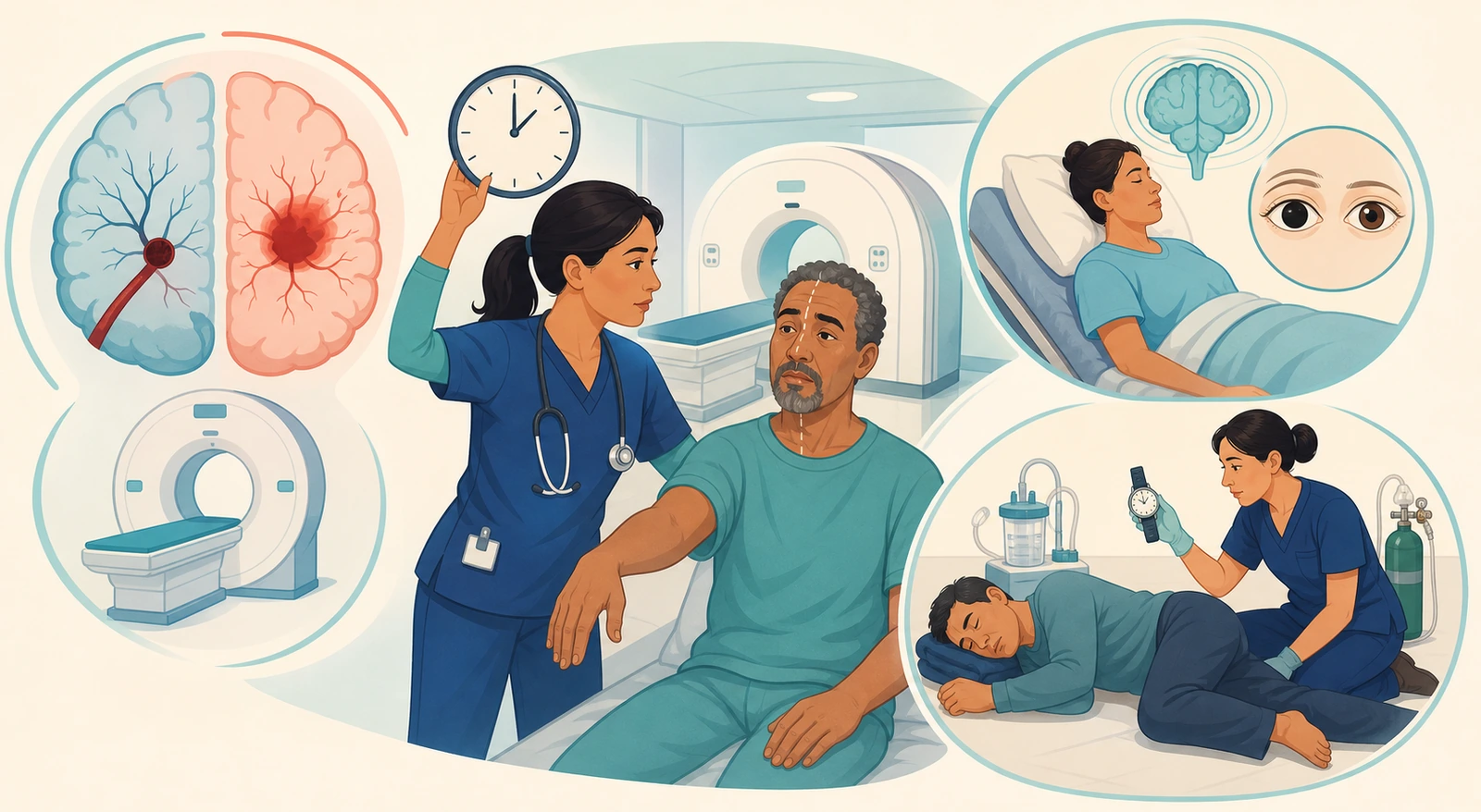
Check airway protection, breathing pattern, oxygenation, and circulation. If there's a sudden neurological deficit, immediately activate your facility's stroke alert or emergency pathway.
Hypoglycemia can mimic unilateral weakness, confusion, or even seizures. Check the blood glucose quickly, but don't delay brain imaging while waiting for the lab draw or other tests.
Confirm the time the patient was last seen normal, not just when the symptoms were discovered. If the patient woke up with symptoms, record both the last known well time before sleep and the discovery time separately.
Establish a baseline by checking consciousness, gaze, pupils, face, arm and leg strength, sensation, language, and articulation in a consistent way.
Prepare for emergency imaging according to your institution's pathway, such as a non-contrast CT. If any changes occur, reassess using the same items and document the time of deterioration.
If you suspect a stroke, do not give any food, water, or oral medications until a swallowing screening is done.
Even if the facial droop looks mild or the patient says "I'm fine," you cannot assume their swallowing function is safe. Verify alternative routes for necessary medications as prescribed and maintain aspiration precautions.
| Cue | Quick Check | Nursing Action |
|---|---|---|
| Balance | Sudden gait unsteadiness, dizziness, loss of coordination | Prevent falls and confirm the onset time and baseline walking status. |
| Eyes | Sudden vision loss, double vision, gaze deviation | Quickly compare left and right visual fields and gaze, and document any changes. |
| Face | One-sided droop when smiling, sensory asymmetry | Compare the face at rest and with instructed movement. |
| Arm | One arm drifting down, decreased grip strength or sensation | Compare both arms in the same position and maintain the emergency pathway as soon as it's detected. |
| Speech | Aphasia, inappropriate words, slurred articulation | Check comprehension, expression, and pronunciation separately, and provide a means of communication. |
| Time | Last known well time and symptom discovery time | Don't estimate. Cross-check with witnesses, records, and device information to document the time down to the minute. |
Don't assume that "treatment is off the table because several hours have passed since symptom onset."
The 2026 AHA/ASA guidelines support IV thrombolysis within 4.5 hours for eligible disabling deficits. They also note that some patients with an unknown onset time or in the 4.5–9 hour window can be selected based on mismatch criteria on advanced imaging. Endovascular therapy for large vessel occlusion can also be considered up to 24 hours, depending on the patient, imaging, and occlusion site. As a nurse, your role is to document the time accurately and not delay imaging or specialist evaluation.
| Category | Ischemic Stroke | Intracerebral Hemorrhage |
|---|---|---|
| Core mechanism | Cerebral blood flow blocked by a thrombus or embolus | Blood accumulates in the brain parenchyma due to vessel rupture |
| Common first steps | ABCs, blood glucose, time last known well, neurological assessment, rapid imaging centered on a non-contrast CT | |
| Main treatment pathway | Alteplase or tenecteplase based on eligibility; evaluate for endovascular therapy if a large vessel occlusion is present | Rapid reversal of anticoagulant effect, smooth and sustained blood pressure management, neurocritical care and neurosurgical evaluation |
| Blood pressure judgment | Targets differ depending on whether thrombolysis or endovascular therapy is given and on reperfusion status | Adjust per institutional protocol, tailored to hemorrhage size and severity and the patient’s condition, while avoiding abrupt fluctuations |
| Contraindication confusion | Do not give a thrombolytic without imaging and eligibility confirmation | Do not give a thrombolytic; share bleeding and anticoagulation information immediately |
| Stage | Changes to observe | What it means and what to do |
|---|---|---|
| Early deterioration | Restlessness, drowsiness, delayed response, headache and vomiting, new confusion | Compare to baseline, report immediately, and repeat the ABCs, blood glucose check, and neurological assessment. |
| Focal signs | Asymmetry in pupil size or reaction, gaze deviation, new unilateral weakness | Report possible brain structure compression or lesion progression, and prepare for emergency imaging and specialist evaluation. |
| Progressive signs | Repeated vomiting, rapid drop in consciousness, abnormal posturing, new seizure | Prepare to protect the airway and manage herniation, and alert the emergency team without delay. |
| Late danger | Cushing’s response: rising systolic blood pressure with widening pulse pressure, bradycardia, irregular breathing | This is a late finding that suggests already-advanced ICP elevation and herniation risk. Don’t wait until all three signs appear together. |
Avoid hypoxia and carbon dioxide accumulation. Prolonged prophylactic hyperventilation is not used because it can reduce cerebral blood flow; temporary hyperventilation during an acute herniation risk is performed only under specialist team direction and monitoring.
Avoid neck rotation and excessive flexion, which can obstruct venous drainage. Unless contraindicated and if hemodynamically tolerated and prescribed, elevate the head of the bed to about 30 degrees.
Cluster necessary care safely, but don’t skip neurological reassessments. Pain, agitation, shivering, fever, vomiting, and excessive suctioning can all raise ICP.
When using hypertonic saline or mannitol, track sodium levels, osmolality, renal function, hemodynamics, and urine output. For an external ventricular drain, maintain the reference height, clamp conditions, and drainage volume exactly as prescribed.
Repeat the GCS or your facility’s neurological tool, pupil check, limb response, and vital signs under the same conditions to document the direction of change and the response to interventions.
If one pupil becomes newly dilated, consciousness drops, and breathing becomes irregular, don’t wait for the “next scheduled assessment.”
Prepare airway equipment and emergency medications, and notify the neurosurgery and neurocritical care teams immediately. Avoid actions that obstruct venous drainage, such as transporting the patient flat or sharply flexing the head.
Immediately note the time the seizure started. Watch for eye and head deviation, where it began, the type of movements, level of consciousness, cyanosis, and any incontinence.
Clear away any dangerous objects nearby and place something soft under the head. If possible, turn the patient onto their side on the floor or a low bed to help secretions drain.
Never hold down the limbs or use restraints. Don't put any objects or your fingers between the teeth, and never give food, water, or oral medications during the convulsion.
Have oxygen and suction ready, but don't force any devices into the mouth during the seizure. If it lasts longer than 5 minutes or starts again without recovery, activate the status epilepticus pathway.
Keep the patient in a side-lying position and check breathing, oxygenation, blood glucose, pupils, muscle strength, any injuries, and recovery time. Hold all oral intake until they are fully awake and can swallow safely.
| Course | Priority Assessment | Next Action |
|---|---|---|
| Brief seizure followed by gradual recovery | Check airway, breathing, any trauma, and postictal confusion | Side-lying position, reassurance, repeat neurological assessments, and accurate documentation |
| Convulsion lasting longer than 5 minutes | Convulsive status epilepticus with risk of treatment delay | Emergency pathway, prescribed benzodiazepine, ABCs, blood glucose, and monitoring |
| Repeated seizures without recovery in between | Emergency: consciousness does not return between seizures | If seizures persist after the first medication, follow institutional guidelines for second-line antiseizure therapy and specialist team evaluation |
| Movements have stopped but consciousness has not returned | Consider nonconvulsive seizure, hemorrhage, or hypoglycemia beyond just a postictal state | ABCs, blood glucose, neurological reassessment, and prepare for continuous EEG and etiology workup |
American Epilepsy Society treatment timeline: The first 0–5 minutes are for emergency stabilization, assessment, and monitoring. If the seizure continues, the 5–20 minute window is the initial treatment phase, which includes benzodiazepines. More than memorizing drug names, the key nursing priority is timing the seizure and making sure the first medication isn't delayed.
Compare the degree of alertness, ability to follow commands, orientation, and GCS components with previous findings.
Document any new changes in left and right pupil size, shape, light reaction, and gaze deviation.
Check for arm drift, grip strength, leg lift, response to painful stimuli, and any left-right differences.
Distinguish changes in comprehension, expression, and articulation. Restrict oral intake until a formal swallowing screening is done.
Link blood pressure values and fluctuations, pulse, oxygenation, and respiratory rate and regularity to neurological changes.
Separately document the onset of changes, time of reporting, timing of imaging/medications/procedures, and the response after each intervention.
The examples below are newly created scenarios for practicing your judgment flow and do not reproduce actual NCLEX questions, answer choices, or correct answers.
The patient was conversing normally until 50 minutes ago, but now their speech is slurred and the right arm drifts downward. Blood glucose is not in the hypoglycemic range, and they are asking for water.
Judgment: Do not give water and activate a stroke alert. Determine the last known well time, check for any anticoagulant use or recent bleeding history, and prepare for a rapid neurological assessment and a non-contrast CT pathway.
The patient woke up at 7 AM and discovered left leg weakness. Before going to sleep at 11 PM, everything was normal.
Judgment: Do not set the onset time as 7 AM. Document 11 PM as the last known well time and 7 AM as the discovery time separately, and proceed without delay to specialist team screening, including advanced imaging.
A patient under observation after a head injury suddenly becomes difficult to arouse, the right pupil is enlarged, the pulse is slowing, and breathing is irregular.
Judgment: Recognize this as a neurological emergency with herniation risk. Support the airway, oxygenation, and hemodynamics, keep the head and neck in a neutral position, and immediately prepare for emergency imaging, prescribed osmotic therapy, and neurosurgical evaluation.
The patient is showing generalized tonic-clonic movements and has not regained consciousness for 6 minutes.
Clinical judgment: Do not restrain them or put anything in their mouth. Position them on their side, protect from injury, prepare oxygen and suction, check blood glucose and monitor. Don’t delay activating the status epilepticus pathway and administering the prescribed benzodiazepine.
After a 90-second seizure, the patient is lying on their side, appears to be sleeping, and responds slowly. The family wants to give water and the usual medications right away.
Clinical judgment: Hold all oral intake until the patient is fully awake and can swallow safely. Check ABCs, blood glucose, pupils, motor strength, and any injuries. Document the seizure type, duration, and recovery course.
1. Did you check airway, breathing, circulation, and blood glucose first?
2. Did you distinguish between the last known well time and the time found?
3. If there’s a sudden focal deficit, did you activate a stroke alert and avoid delaying the CT?
4. Did you hold oral intake before a swallow screen?
5. Did you avoid assuming ischemic vs. hemorrhagic based on symptoms alone?
6. Did you confirm that blood pressure targets differ for IVT, EVT, and ICH?
7. Did you track trends in consciousness, pupils, motor function, and breathing to catch worsening ICP?
8. Did you time the seizure onset and maintain protection, side-lying positioning, and prohibited actions?
9. If the seizure lasted over 5 minutes or recurred without recovery, did you activate the emergency pathway?
10. After the intervention, did you reassess using the same neurological items?
Official standards: NCSBN, 2026 NCLEX-RN Test Plan · American Heart Association/American Stroke Association, 2026 Acute Ischemic Stroke Guideline · American Heart Association/American Stroke Association, 2022 Spontaneous Intracerebral Hemorrhage Guideline · Brain Trauma Foundation, Guidelines for the Management of Severe TBI, 4th Edition · American Epilepsy Society, Guideline for Treatment of Prolonged Seizures · CDC, First Aid for Seizures
This independent study flow is built on the 2026 AHA/ASA time-, imaging-, and indication-based ischemic stroke care, the 2022 intracerebral hemorrhage management, official ICP and seizure emergency guidelines, and the NCSBN clinical judgment scope. In actual practice, follow the latest prescriptions, your institution’s stroke, ICP, and status epilepticus protocols, and patient-specific contraindications.
Content boundary: The local feedback material only identified recurring study topics. No actual exam items, answer choices, correct answers, tables, screens, patient values, or illustrations were copied or recreated. All example situations, sentences, tables, judgment sequences, and images were newly developed.
다음 이론을 계속 학습하려면 로그인하세요.
로그인하고 계속 학습필기노트, 하이라이터, 메모는 잘 쓰고 있어?
내보내줘운영진이 검토할게요!
마이페이지에서 차단한 회원을 관리할 수 있어요.