컨텐츠 내용을 수정할 수 있습니다
Don't just focus on drainage output or SpO₂ alone. Learn to quickly distinguish whether sudden respiratory distress is coming from the patient, the chest tube system, the pulmonary vessels, or ventilation.
Core Goal: Assess the patient first → Check the chest tube and drainage unit → Differentiate pneumothorax, pulmonary embolism, and respiratory failure → Report and support immediately → Re-evaluate the same items — follow this sequence for your clinical judgment.
A chest tube is a drainage device, but if you only stare at the equipment, you might miss a tension pneumothorax or respiratory fatigue. Conversely, clamping or stripping the tube just because bubbling or drainage has stopped can be dangerous. Always look at the patient's condition and the entire system together.
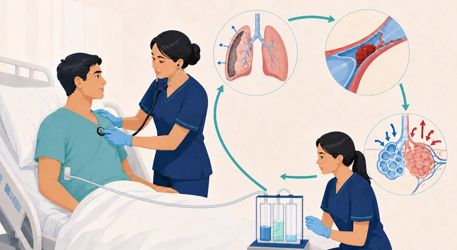
Check responsiveness, airway patency, ability to speak, and any secretions or aspiration risk.
Compare respiratory rate and depth, accessory muscle use, bilateral chest movement and breath sounds, skin color, and signs of respiratory fatigue.
Check the SpO₂ waveform and trend, and verify the prescribed oxygen is actually connected, but don't just trust a number that looks normal.
Check pulse, blood pressure, skin perfusion, chest pain, jugular vein distention, and any new signs of shock.
After that, look at the chest tube insertion site, tubing path, drainage unit position, water-seal fluctuation/bubbling, and drainage trends.
If you find unilateral loss of breath sounds, sudden severe dyspnea, hypotension, cyanosis, and decreased level of consciousness together, suspect tension pneumothorax first.
Call the emergency team immediately and prepare oxygen, monitoring, decompression, and chest tube management. Do not leave the patient alone while waiting for test results, and never arbitrarily clamp the chest tube without knowing the cause.
| Area to Check | What It Shows | Nursing Judgment |
|---|---|---|
| Collection chamber | Where drainage like blood or exudate collects, besides air | Mark the amount, color, consistency, and hourly trends; connect any sudden increase or decrease to the patient's condition |
| Water-seal chamber | A one-way barrier that lets air escape but prevents it from re-entering the pleural space | Check fluctuation with breathing and the timing/duration of bubbling to differentiate between recovery, blockage, or an air leak |
| Suction chamber | Regulates the prescribed negative pressure based on the system type | Distinguish between wet and dry systems; verify the prescribed setting, wall suction connection, and device instructions together |
| Insertion site | Where the tube is secured to the chest wall and protected by a dressing | Check for securement, drainage/bleeding, infection, subcutaneous emphysema, new pain, and any change in the external tube length |
Basic positioning: Always keep the drainage unit below the insertion site and upright. Make sure the tubing isn't compressed, kinked, or lying under the patient. Avoid long dependent loops on the floor and reduce tension. Even when moving the patient, don't lift the drainage unit onto the bed or routinely clamp the tube.
In the early stage of a pneumothorax, air may escape during coughing or exhalation. Rather than the bubbling itself, watch the patient's breathing and whether it decreases over time.
New or persistent bubbling can suggest an air leak on the patient side or at a connection. Check systematically, starting from the patient and insertion site, then the connections, and finally the drainage unit.
This can happen due to lung re-expansion, but also consider tube kinking, blockage, or a dependent fluid-filled loop. Don't assume improvement based solely on the cessation of drainage.
If this occurs alongside worsening patient breathing, tube displacement, or a sudden gush or stop of drainage, get help immediately and correct the cause.
Don't use "If you see bubbling, clamp the tube to find the leak" as a general rule.
The BTS clinical statement limits clamping a bubbling chest tube to specific situations under the supervision of a specialist pleural team. Arbitrary clamping during a persistent air leak can create a risk of tension pneumothorax, so follow the prescription, device instructions, and your institutional protocol.
Reassess the patient's breathing and breath sounds first, then check for kinks, compression, dependent loops, and connections. Do not strip or forcefully milk the tube to move contents without a prescription.
Immediately record the amount and color, and check blood pressure, pulse, skin perfusion, pain, and the dressing. New bright red drainage or hemodynamic changes suggest possible bleeding and must be reported right away.
Do not place the contaminated end on the floor and call for help immediately. Restore a sterile connection or set up a new system according to your institution's chest tube emergency protocol and device guidelines, and do not clamp the tube for an extended period without an order.
Do not push it back in. Call for emergency help right away, apply a sterile occlusive dressing per your institutional policy, and continuously monitor for respiratory distress, unilateral breath sounds, and signs of tension pneumothorax.
Check the patient's condition, assess the system for damage and whether the water seal is maintained, and set the unit upright. If the water seal is compromised or contaminated, replace it with a new unit according to the guidelines.
| Timing | Key Nursing Care | Changes to Suspect Immediately |
|---|---|---|
| Pre-procedure | Verify consent, labs, and anticoagulation-related instructions; document baseline vital signs, SpO₂, breath sounds, and dyspnea | Inability to cooperate, unstable respiratory/circulatory status, bleeding risk, and difficulty maintaining position |
| Intra-procedure | Position the patient upright with arms supported if possible, assist with aseptic technique, and prevent sudden movements | Chest pain, persistent cough, worsening dyspnea, dizziness, and hypotension |
| Post-procedure | Compare respiratory rate, SpO₂, bilateral breath sounds, puncture site, pain, and patient response to baseline | New unilateral diminished breath sounds, sudden dyspnea, chest pain, and hypoxemia raise concern for complications like pneumothorax |
| Clue | What to Check | Key Judgment Point |
|---|---|---|
| Sudden symptoms | Unexplained dyspnea, pleuritic chest pain, tachycardia, hypoxemia, dizziness/syncope, hemoptysis | Clear breath sounds do not rule out PE; evaluate alongside other potential causes |
| Thrombotic risk | Recent surgery, trauma, hospitalization, prolonged immobility, cancer, pregnancy/postpartum, estrogen use, prior VTE | First, organize the risk factors and clinical probability, then decide on the diagnostic pathway |
| DVT clues | New unilateral leg swelling, pain, warmth, or circumference difference | Do not massage the leg; report immediately to prepare for the diagnostic and anticoagulation pathway |
| High-risk changes | Persistent or recurrent hypotension, shock, decreased level of consciousness, severe hypoxemia/ventilatory failure, right ventricular strain | Simultaneously prepare for emergency/critical care and the possibility of advanced reperfusion therapy |
D-dimer is not a confirmatory test for PE. In patients with low or intermediate clinical probability, a negative result can help reduce the need for imaging, but if the probability is high or the result is elevated, it should lead to an appropriate imaging pathway such as CTPA. A single value alone does not confirm or rule out PE.
This is when oxygen does not move adequately from the alveoli into the blood. Connect SpO₂, PaO₂, oxygen requirements, breath sounds, and imaging findings to the underlying cause.
This is when the body cannot eliminate enough CO₂. Shallow, slow breathing, drowsiness/confusion, headache, and changes in PaCO₂ and pH are key indicators.
In COPD exacerbation, severe asthma, neuromuscular fatigue, sedation/overdose, and worsening lung disease, both problems can overlap.
When supplemental oxygen is given, SpO₂ can be maintained even as ventilatory failure and rising CO₂ progress. Always assess level of consciousness, depth of breathing, and ABG results together.
If a patient who was breathing heavily suddenly becomes quiet and their respiratory rate and effort drop, think fatigue first — not improvement.
Shallow breathing, inability to speak, drowsiness or confusion, weak cough, silent chest, and worsening acidosis are all signs of impending ventilatory failure. Call for senior help immediately and prepare for airway and ventilation support.
The examples below are newly created scenarios to practice your clinical reasoning flow and do not reproduce actual NCLEX items, answer choices, or correct answers.
A patient with a chest tube for a pneumothorax now has continuous bubbling in the water-seal chamber that wasn’t there before. The patient is awake, and their SpO₂ and breathing are similar to baseline.
Judgment: Don’t clamp the tube just because you see bubbling. Check the patient and the insertion site first. Then inspect the tubing from the patient toward the drainage unit for kinks, disconnections, or loose connections, and report the new air leak to the provider.
After pleural fluid drainage, the patient suddenly complains of chest pain and dyspnea, and breath sounds are decreased on the side of the procedure.
Judgment: Suspect a post-procedure pneumothorax first. Immediately reassess oxygenation and hemodynamics, and call for emergency help while preparing for imaging, decompression, and chest tube insertion.
A patient who had limited mobility after recent surgery develops sudden dyspnea and pleuritic chest pain, then briefly loses consciousness, and their blood pressure drops.
Judgment: Suspect cardiopulmonary decompensation from a high-risk acute PE. Activate the emergency team immediately to prepare oxygen, monitoring, IV access, and the diagnostic pathway. Support decisions for risk-appropriate anticoagulation and reperfusion therapy.
A patient who had severe tachypnea and accessory muscle use gradually starts breathing shallowly and slowly, becomes drowsy, and their SpO₂ is maintained on supplemental oxygen.
Judgment: Don’t interpret this as improvement. Suspect ventilatory fatigue and CO₂ retention. Immediately assess the airway, breathing, level of consciousness, and ABG, and report so a rapid decision can be made about the suitability of NIV or the need for intubation and mechanical ventilation.
1. Is the patient talking, awake, and protecting their airway?
2. Is chest wall movement and breath sounds symmetric bilaterally, and are blood pressure and perfusion maintained?
3. Is the drainage unit below the insertion site, upright, and are the tubes free of kinks?
4. In what context did the bubbling, tidaling, or drainage changes appear relative to the patient’s condition?
5. Are you avoiding clamping or stripping the chest tube without an order?
6. After a thoracentesis, is there new chest pain, dyspnea, or a unilateral decrease in breath sounds?
7. Are there PE risk factors along with hypotension, syncope, hypoxia, or right heart strain?
8. Have you separated oxygenation from ventilation and looked at SpO₂, consciousness, and ABG together?
9. After an intervention, have you rechecked the same parameters, and is escalation of care not being delayed?
Official Sources: NCSBN, 2026 NCLEX-RN Test Plan · British Thoracic Society, Clinical Statement on Pleural Procedures · ACC/AHA, 2026 Acute Pulmonary Embolism Guideline Summary · ERS/ATS, Noninvasive Ventilation for Acute Respiratory Failure
I’ve put together this self-study guide based on the NCSBN’s drainage device, thoracentesis, ventilator, and impaired ventilation/oxygenation activities, the BTS’s pleural procedure and chest tube safety principles, the 2026 ACC/AHA guidelines on acute PE diagnosis and risk classification, and the ERS/ATS principles for NIV patient selection and early reassessment. In actual practice, always follow the latest guidelines, prescriptions, device manuals, and your facility’s protocols.
This material is an educational summary I independently reorganized around topics that came up repeatedly in local feedback. It does not restore or reproduce actual NCLEX questions, answers, answer choices, exam screens, original tables, patient materials, or source images. In real patient care, please follow the latest clinical guidelines, institutional protocols, and the judgment of the responsible healthcare team.
다음 이론을 계속 학습하려면 로그인하세요.
로그인하고 계속 학습필기노트, 하이라이터, 메모는 잘 쓰고 있어?
내보내줘운영진이 검토할게요!
마이페이지에서 차단한 회원을 관리할 수 있어요.