컨텐츠 내용을 수정할 수 있습니다
Instead of focusing on just one finding like edema or low blood pressure, learn to separate congestion from hypoperfusion and master a decision-making sequence so you don't miss cardiogenic shock or emergencies involving the extremities and pulmonary vessels.
Core Objective: Think through this flow: Is there congestion? → Is organ perfusion (hypoperfusion) dropping? → What is the underlying cause? → Is the patient improving after the intervention?
A heart failure patient can have pulmonary congestion and hypoperfusion at the same time, and the direction of treatment—fluids, vasoactive agents, or mechanical support—changes depending on the cause of shock. Don't choose an intervention based on a single vital sign or a disease name alone.
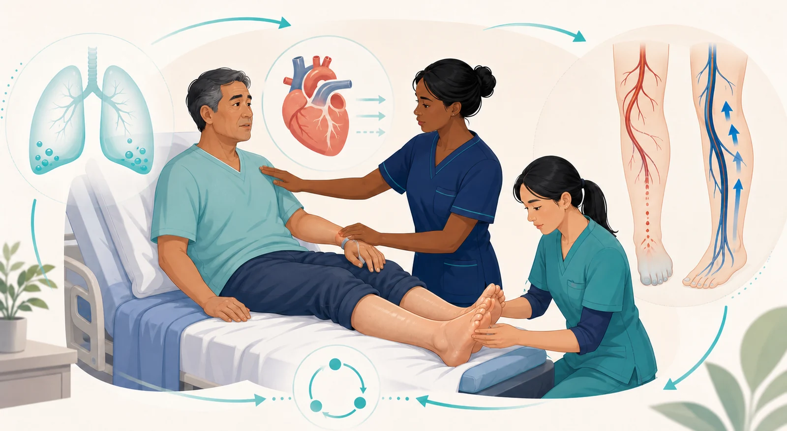
Check if the airway is open and whether respiratory distress, hypoxemia, or fatigue is progressing.
Look for new-onset confusion, anxiety, lethargy, and skin that is cool and clammy or abnormally warm.
Connect the dots between BP trends, pulse strength, skin color/temperature, and capillary refill.
Check if a drop in urine output and changes in renal function suggest decreased organ perfusion.
Reassess the direction of symptom, vital sign, and lab changes rather than relying on a single-point value.
| Assessment Axis | Key Clues | Connected Clinical Judgment |
|---|---|---|
| Pulmonary Congestion | Dyspnea, orthopnea, paroxysmal nocturnal dyspnea, crackles, decreased oxygenation | Respiratory burden and whether pulmonary edema is progressing; need for immediate respiratory support |
| Systemic Congestion | Jugular venous distention, peripheral edema, abdominal bloating, rapid weight gain over a short period | Trend of fluid accumulation and response to treatment; risk for skin breakdown |
| Hypoperfusion | Fatigue, cool skin, weak pulses, decreased urine output, confusion, hypotension | Decreased cardiac output and worsening organ function; potential for cardiogenic shock |
| Treatment Response | Changes in breathing, BP, level of consciousness, urine output, edema, and weight | Repeatedly check if the intervention is reducing congestion while maintaining perfusion |
Don't just memorize 'left-sided failure affects the lungs, right-sided failure affects the body' and stop there. In a real patient, findings from both sides can overlap. Look at the whole picture: is there clear pulmonary congestion along with JVD, edema, and hypoperfusion at the same time?
Assess responsiveness, airway, work of breathing, oxygenation, pulses, BP trends, and skin and urine status.
Place the patient in a semi-Fowler's or as upright a position as tolerated and begin monitoring. If hypoxemia or respiratory failure is present, connect them to oxygen and respiratory support per institutional protocols.
Activate a rapid response or emergency team for progressive dyspnea, a new change in level of consciousness, chest discomfort, or severe hypoperfusion.
Prepare for assessments per orders/protocols, such as an ECG, IV access, lab work, chest imaging, and ultrasound. Identify triggers like ischemia, arrhythmias, infection, or medication/dietary non-adherence.
Repeatedly check if breathing, oxygenation, BP, pulse, level of consciousness, urine output, skin temperature, and signs of congestion are actually improving.
If you see hypotension that could be from pulmonary congestion or cardiogenic shock, don't automatically give a big fluid bolus to every patient.
Fluid decisions should be based on the cause, preload, signs of congestion, and how the patient responds. In a patient whose heart pump is failing, careless fluid loading can worsen pulmonary congestion, so notify the medical and emergency teams right away and follow the cause-specific treatment pathway.
Circulating volume is low from hemorrhage or fluid loss. You'll see tachycardia, weak pulses, cool clammy skin, narrowing pulse pressure, and hypoperfusion clues — but also look for the bleeding source and the cause of the loss.
Vasodilation and maldistribution are the core problem. In early septic shock, the skin can actually be warm, so don't assume "warm = not shock." Assess the infection source and organ perfusion together.
Forward blood flow is inadequate because the pump is failing. In the setting of myocardial ischemia or heart failure, you may see hypoperfusion along with pulmonary and systemic congestion.
There's a mechanical blockage to blood flow — think pulmonary embolism, cardiac tamponade, or tension pneumothorax. Removing the obstruction is key, so report any sudden respiratory or circulatory deterioration immediately.
Don't apply the same fluid formula and the same positioning to every type of shock.
Connect the cause, respiratory status, congestion, cardiac function, bleeding risk, and the response to interventions. The textbook categories are a starting point; actual treatment follows current guidelines, institutional protocols, and provider orders.
| Condition | Typical clues | Nursing judgment |
|---|---|---|
| Peripheral Arterial Disease (PAD) | Pain with walking/claudication, cool pale skin, weak pulses, foot wounds that don't heal well | Compare bilateral pulses, skin, and sensation; teach foot protection, smoking cessation, prescribed structured exercise, and vascular risk management |
| Chronic Venous Insufficiency (CVI) | Edema and heaviness, venous skin changes, worse with prolonged standing, pulses usually palpable | After confirming arterial status and the prescription, apply elevation and compression pathways, and prevent skin breakdown |
| Deep Vein Thrombosis (DVT) | Usually unilateral swelling, pain/tenderness, warmth, redness | Don't rely on clinical findings alone to rule it in or out — report immediately to connect to diagnostic evaluation and the anticoagulation treatment pathway |
Sudden dyspnea, pleuritic chest pain, hemoptysis, syncope, or unexplained hypoxemia — think pulmonary embolism first.
Don't massage a leg where you suspect DVT, and don't try to confirm it with Homan's sign. Immediately assess the patient's breathing and circulation, and activate the emergency response pathway.
Sudden onset of pain, pallor, pulselessness, coolness, paresthesia, and weakness suggests blood flow to the limb has been abruptly cut off. Don't wait for every finding to be complete — request an urgent vascular evaluation right away.
Compare pulses or Doppler signals, skin color and temperature, and capillary refill with the opposite limb.
Numbness, tingling, or decreased toe movement can be a warning of advanced ischemia.
If pulses, temperature, color, pain, movement, or sensation suddenly worsen after revascularization, report immediately.
Don't apply pressure or massage a symptomatic limb on your own, and don't apply direct heat without an order.
Use the same scale every day, ideally in the morning after you empty your bladder and wearing similar clothes, so you can track the trend of any changes.
Record shortness of breath, discomfort when lying down, swelling, changes in your sleep, fatigue, appetite, and medication use right alongside your weight changes.
Follow the weight and symptom warning thresholds your healthcare team has set for you. If you notice rapid worsening or new breathing difficulty, don't wait for your next scheduled appointment.
If you have PAD, look for any sores, blisters, color changes, coolness, or decreased sensation. Avoid going barefoot, using excessive heat, or wearing shoes that don't fit well.
The examples below are newly created scenarios to practice your judgment flow and do not reproduce actual NCLEX questions, answer choices, or correct answers.
A heart failure patient says it's hard to lie flat, and you notice new crackles, cool hands, a weak pulse, and decreased urine output.
Judgment: First, consider the possibility of cardiogenic shock, where pulmonary congestion and low perfusion are happening at the same time. Raise the head of the bed, immediately reassess breathing, oxygenation, and perfusion, and activate the emergency team. Avoid the path of automatically giving a large fluid bolus just because you see low blood pressure.
After a revascularization procedure, one foot becomes colder and paler than the other, a previously palpable pulse becomes weaker, and the patient feels tingling.
Judgment: Don't wait, thinking it's normal recovery pain. Compare pulses or Doppler signals, skin color and temperature, and sensation and movement on both sides, and report your findings to the vascular team immediately.
A patient who has had pain and swelling in one calf for a few days suddenly complains of shortness of breath and chest pain.
Judgment: An emergency assessment for pulmonary embolism takes priority over relieving the leg pain. Quickly check breathing and circulation, call for an emergency response, and do not massage the leg.
1. What is the most immediate threat right now — breathing, circulation, or both?
2. Did I identify the clues for congestion and the clues for low perfusion separately?
3. Did I look at the trend in mental status, skin, pulses, and urine output, rather than just one blood pressure reading?
4. Which type of shock does the cause most closely resemble — hypovolemic, distributive, cardiogenic, or obstructive?
5. When heart failure and low blood pressure occur together, did I avoid giving fluids indiscriminately?
6. Did I differentiate the leg problem as arterial insufficiency, venous insufficiency, or DVT?
7. Did I miss any warning signs of sudden limb ischemia or a pulmonary embolism?
8. After the intervention, did I reassess the same perfusion parameters?
Official Standards: NCSBN, 2026 NCLEX-RN Test Plan · AHA/ACC/HFSA, 2022 Heart Failure Guideline · American Heart Association, Heart Failure Warning Signs and Physical Changes to Report · American College of Cardiology, 2025 Cardiogenic Shock Guidance · ACC/AHA, 2024 Peripheral Artery Disease Guideline · American Society of Hematology, VTE Treatment Guidelines
This patient-first, independent learning flow was structured based on the NCSBN's categories of hemodynamics, decreased cardiac output, tissue perfusion, and emergency situations, along with official guidelines for heart failure, cardiogenic shock, PAD, and VTE. Since medication, fluid, and device settings vary depending on the patient's condition, the latest guidelines, and institutional protocols, they are not presented as fixed prescriptions in this summary.
This material is an educational summary independently reconstructed from repeated learning topics in local feedback. It does not restore or reproduce actual NCLEX questions, answers, options, test screens, original tables, patient data, or source images. In actual patient care, follow the latest clinical guidelines, institutional protocols, and the judgment of the attending healthcare team.
다음 이론을 계속 학습하려면 로그인하세요.
로그인하고 계속 학습필기노트, 하이라이터, 메모는 잘 쓰고 있어?
내보내줘운영진이 검토할게요!
마이페이지에서 차단한 회원을 관리할 수 있어요.